Article added / Artikel hinzugefügt 01.10.2021
Generally Articles and Discussions about Osteosarcoma in Dogs
→ Evaluations of phylogenetic proximity in a group of 67 dogs with
osteosarcoma: a pilot study
Article added / Artikel hinzugefügt 01.10.2021
Generally Articles and Discussions about Osteosarcoma in Dogs
→ Canine Periosteal Osteosarcoma
Images added / Abbildungen hinzugefügt 02.05.2019
Generally Sonography Atlas of Dogs →
Cardiovascular system → Pulmonary vessels
New subcategory added / Neue Unterkategorie hinzugefügt 02.05.2019
Generally Sonography Atlas of Dogs →
Cardiovascular system → Pulmonary vessels
Images added / Abbildungen hinzugefügt 01.05.2019
Generally Sonography Atlas of Dogs →
Cardiovascular system → Heart valvular diseases


Generally Sonography Atlas of Dogs
(Allgemeiner Sonographie-Atlas von Hunden)
Muscoskeletal System - Knee
(Muskoskelettales System - Knie)
Imagen ecográfica correspondient e al corte sagital de la región suprarrotuliana en la que se visualiza el tendón del músculo cuádriceps femoral con estructura fibrilar y peritendón hiperecogénico (flechas). P: proximal, D: distal, TF: tróclea del fémur.
Ultrasound image correspondient and the sagittal section of the region suprapatellar in which the tendon of the quadriceps femoris muscle displayed c on peritendon hyperechoic fibrillar structure ( arrows). Q: proximal , D : distal , TF : trochlea of the femur.
Soler Laguía, Marta
"Diagnóstico por imagen de la articulación de la rodilla en la especie canina"
oai:digitum.um.es:10201/40906

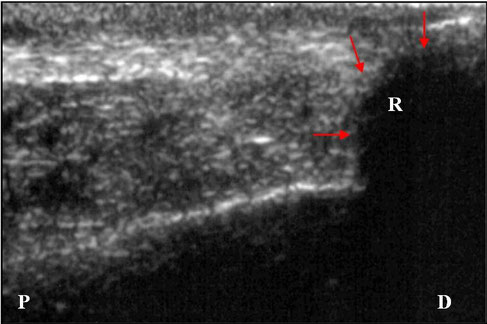
Imagen ecográfica del corte sagital de la regi
ón suprarrotuliana en la que se visualiza la superficie
convexa hiperecogénica de la rótula (flechas). P: proximal, D: distal, R: rótula.
Sagittal ultrasound image of the region suprapatellar ng
displayed in the hyperechoic convex surface of the patella ( arrows). Q: proximal , D : distal , R :
kneecap.
Soler Laguía, Marta
"Diagnóstico por imagen de la articulación de la rodilla en la especie canina"
oai:digitum.um.es:10201/40906

Imagen ecográfica del corte sagital de la región
suprarrotuliana en la que visualizamos el cartílago articular de la tróclea del fémur, como una línea hipoecogénica lisa
demarcada por dos interfases
hiperecogénicas (flechas). P: proximal, D: distal, R: rótula, F: fémur.
Sagittal ultrasound image of the resuprapatellar region in
which we visualize the articular cartilage of the trochlea
l femur, as a hypoechoic smooth line demarcated by two interfaces hyperechoic ( arrows). Q: proximal , D : distal , R : patella, F: femur.
Soler Laguía, Marta
"Diagnóstico por imagen de la articulación de la rodilla en la especie canina"
oai:digitum.um.es:10201/40906

Imagen ecográfica correspondiente al corte sagital del ligamento rotuliano, que se visualiza con estructura fibrilar y peritendón
hiperecogénico (flechas). P: proximal, D: distal, R: rótula, TF: tróclea del fémur.
Ultrasound image corresponding to the sagittal section of the patellar ligament, which is displayed with hyperechoic fibrillar
structure and peritendon ( arrows). Q: proximal , D: distal, R : patella TF: trochlea of the
femur.
Soler Laguía, Marta
"Diagnóstico por imagen de la articulación de la rodilla en la especie canina"
oai:digitum.um.es:10201/40906

Imagen ecográfica del corte sagital de la región infrarrotuliana en la que se observa el cuerpo adiposo infrarrotuliano, caudal al
ligamento rotuliano con una ecogenicidad media homogénea (flechas). P: proximal, D: distal, F: fémur, T: tibia, LR: ligamento rotuliano.
Sagittal ultrasound image of the infrapatellar region where the infrapatellar fat pad is observed flow to the patellar ligament
with a homogeneous medium echogenicity ( arrows). Q: proximal , D : distal , F: femur, T: warm , LR : patellar ligament.
Soler Laguía, Marta
"Diagnóstico por imagen de la articulación de la rodilla en la especie canina"
oai:digitum.um.es:10201/40906

Imagen ecográfica de un corte longitudinal sagital de la región infrarrotuliana en la que se visualiza el ligamento cruzado
craneal (flechas) extendiéndose desde el área intercondilar central de la tibia hasta la fosa intercondilar del fémur. P: proximal, D: distal, F: fémur, T: tibia.
Ultrasound image of a sagittal Slitting infrapatellar region where the cranial cruciate ligament ( arrows ) extending from the
central intercondylar area of the tibia to the intercondylar fossa of the femur is displayed . Q:proximal , D : distal , F:
femur, T: tibia.
Soler Laguía, Marta
"Diagnóstico por imagen de la articulación de la rodilla en la especie canina"
oai:digitum.um.es:10201/40906

Imagen ecográfica correspondiente a un corte sagital de la
región infrarrotuliana en la que se pueden observar los dos ligamentos cruzados craneal (flecha azul) y caudal
(fl
echa amarilla) formando una “V” y presentando la misma ecogenicidad. P: proximal, D: distal, F: fémur, T: tibia.
Corresponding ultrasound image a sagittal section of the
region infrapatellar where you can see the two cross head (blue arrow ) ligaments and flow (fl yellow arrow) forming a " V " and presenting the same echogenicity. P :
proximal , D : distal , F: femur, T: tibia.
Soler Laguía, Marta
"Diagnóstico por imagen de la articulación de la rodilla en la especie canina"
oai:digitum.um.es:10201/40906

Imagen ecográfica de un corte sagital de la región infrarrotuliana en la que se visualiza el ligamento cruzado caudal, desde el
punto de fijación de la superficie lateral del cóndilo femoral medial, dirigiéndose paralelo a éste, hacia la escotadura poplítea de la tibia (flecha). P: proximal, D: distal, F: fémur, T: tibia.
Ultrasound image of a sagittal section of the infrapatellar region where the cruciate ligament is displayed flow , from the point
of attachment of the side surface of the medial femoral condyle , addressing parallel thereto , to the pop recess Litea
tibia ( arrow ) . Q: proximal , D : distal , F: femur, T: tibia.
Soler Laguía, Marta
"Diagnóstico por imagen de la articulación de la rodilla en la especie canina"
oai:digitum.um.es:10201/40906

Corte sagital de la región infrarrotuliana en la que se observa el cartílago articular del cóndilo lateral del fémur
hipoecogénico, demarcado por dos líneas hiperecogénicas lisas, paralelas a la superficie del cóndilo (flechas). P: proximal, D: distal, F: fémur.
Sagittal section of the infrapatellar region in which articular cartilage is observed lateral condyle of hypoechoic femur ,
demarcated by two parallel to the surface of the condyle (arrows) hyperechoic smooth lines . Q: proximal , D : distal , F: femur.
Soler Laguía, Marta
"Diagnóstico por imagen de la articulación de la rodilla en la especie canina"
oai:digitum.um.es:10201/40906

Imagen ecográfica del corte sagital de la región lateral en la que se visualiza el tendón de origen del músculo extensor digital
largo delimitado por el peritendón hiperecogénico (flechas azules), localizándose inmediatamente
lateral y superficial al menisco lateral (flechas rojas). P: proximal, D: distal, F: fémur, T:
tibia.
Sagittal ultrasound image of the lateral region in which the tendon origin of extensor digitorum longus muscle delimited by the
hyperechogenic peritendon (blue arrows) is displayed ,being located immediately latera
lateral meniscus surface to ly ( red arrows) . Q: proximal , D : distal , F: femur, T: tibia.
Soler Laguía, Marta
"Diagnóstico por imagen de la articulación de la rodilla en la especie canina"
oai:digitum.um.es:10201/40906

Imagen ecográfica correspondiente al corte sagital de la región medial en la que observamos el menisco medial como una estructura
hipoecogénica homogénea con forma triangular (flechas). P: proximal, D: distal, F: fémur, T:
tibia.
Ultrasound image corresponding to the sagittal section of the medial region in which we observe the medial meniscus as a
homogeneous hypoechoic triangular structure ( arrows). P :proximal , D : distal , F: femur, T:
tibia.
Soler Laguía, Marta
"Diagnóstico por imagen de la articulación de la rodilla en la especie canina"
oai:digitum.um.es:10201/40906

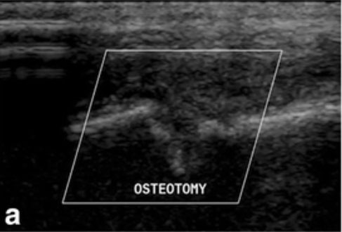
Three ultrasonographic images made of the osteotomy site of a dog.
(a) 1 month postoperatively, (b) 2 months postoperatively; (c) 3 months postoperatively. Note the presence of strongly reflective, irregular interfaces within the osteotomy gap indicative of bone production, and compatible with healing of the osteotomy in all images.
Marije RisseladaEmail author, Matthew D. Winter, Daniel D. Lewis, Emily Griffith, Antonio Pozzi: "Comparison of three imaging modalities used to evaluate bone healing after tibial tuberosity advancement in cranial cruciate ligament-deficient dogs and comparison of the effect of a gelatinous matrix and a demineralized bone matrix mix on bone healing – a pilot study". BMC Vet Res (2018) 14: 164. https://doi.org/10.1186/s12917-018-1490-4

Three ultrasonographic images made of the osteotomy site of a dog.
(a) 1 month postoperatively, (b) 2 months postoperatively; (c) 3 months postoperatively. Note the presence of strongly reflective, smooth, concave (hourglass) interfaces within the osteotomy gap indicative of bone production, and compatible with healing of the osteotomy in all images.
Marije RisseladaEmail author, Matthew D. Winter, Daniel D. Lewis, Emily Griffith, Antonio Pozzi: "Comparison of three imaging modalities used to evaluate bone healing after tibial tuberosity advancement in cranial cruciate ligament-deficient dogs and comparison of the effect of a gelatinous matrix and a demineralized bone matrix mix on bone healing – a pilot study".

Diese Webseite wurde mit Jimdo erstellt! Jetzt kostenlos registrieren auf https://de.jimdo.com


