

Article added / Artikel hinzugefügt 01.10.2021
Generally Articles and Discussions about Osteosarcoma in Dogs
→ Evaluations of phylogenetic proximity in a group of 67 dogs with
osteosarcoma: a pilot study
Article added / Artikel hinzugefügt 01.10.2021
Generally Articles and Discussions about Osteosarcoma in Dogs
→ Canine Periosteal Osteosarcoma
Images added / Abbildungen hinzugefügt 02.05.2019
Generally Sonography Atlas of Dogs →
Cardiovascular system → Pulmonary vessels
New subcategory added / Neue Unterkategorie hinzugefügt 02.05.2019
Generally Sonography Atlas of Dogs →
Cardiovascular system → Pulmonary vessels
Images added / Abbildungen hinzugefügt 01.05.2019
Generally Sonography Atlas of Dogs →
Cardiovascular system → Heart valvular diseases


Generally Sonography Atlas of Dogs
(Allgemeiner Sonographie-Atlas von Hunden)
Muscoskeletal System - Elbow
(Muskoskelettales System - Ellbogengelenk)

Primary flexor enthesopathy. Medial (A) and dorsal (B) views of the elbow of a 14 month-old Golden retriever dog produced with computed tomographic (CT) volume-rendering, with the overlying transverse and longitudinal planes used to produce the sonographc images on the right (respectively). Large (M) and small (m) mineral fragments are depicted in all images. These mineral bodies are embedded in a heterogeneous mass of moderate echogenicity (arrowheads) consistent with fibrous and metaplastic soft tissue, in part confluent with some of the flexor tendons (T). Note the stronger acoustic enhancement associated with the larger mineral bodies.
With special thanks to the authors of the book "Small Animal Ultrasonography" , Marc-André d’Anjou and Dominique Penninck

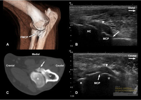
Fragmentation of the medial coronoid process. A: Medial view of the elbow of a 12 month-old Labrador retriever dog produced with computed tomographic (CT) volume-rendering, on which the fragmentation of the medial coronoid process (FMCP) is well visualized. B: This longitudinal sonographic image was obtained with the probe placed medially on the joint, the marker oriented proximally. The joint capsule is distended and thickened (arrowhead) and a gap (arrow) is present at the distal aspect of the medial coronoid process (MCP). The humeral condyle (HC) is mildly irregular. C and D: Corresponding transverse CT (C) and sonographic (D) images in which a fissure (arrows) extends through the MCP. The joint capsule is distended (arrowhead). R, radial head. U, ulna.
With special thanks to the authors of the book "Small Animal Ultrasonography" , Marc-André d’Anjou and Dominique Penninck
