Article added / Artikel hinzugefügt 01.10.2021
Generally Articles and Discussions about Osteosarcoma in Dogs
→ Evaluations of phylogenetic proximity in a group of 67 dogs with
osteosarcoma: a pilot study
Article added / Artikel hinzugefügt 01.10.2021
Generally Articles and Discussions about Osteosarcoma in Dogs
→ Canine Periosteal Osteosarcoma
Images added / Abbildungen hinzugefügt 02.05.2019
Generally Sonography Atlas of Dogs →
Cardiovascular system → Pulmonary vessels
New subcategory added / Neue Unterkategorie hinzugefügt 02.05.2019
Generally Sonography Atlas of Dogs →
Cardiovascular system → Pulmonary vessels
Images added / Abbildungen hinzugefügt 01.05.2019
Generally Sonography Atlas of Dogs →
Cardiovascular system → Heart valvular diseases


Generally Sonography Atlas of Dogs
(Allgemeiner Sonographie-Atlas von Hunden)
Muscoskeletal System - Hip joint
(Muskoskelettales System - Hüftgelenk)
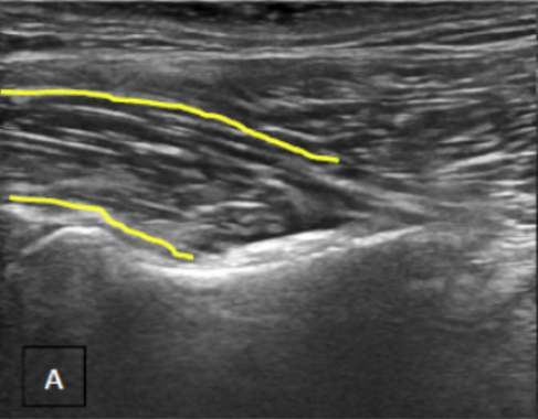
Evaluation of a normal iliopsoas tendon. (a) Longitudinal and (b) cross-sectional views of the tendon of insertion onto the lesser trochanter. (c) A longitudinal view of the tendon of insertion. (d) A longitudinal view of the iliopsoas muscle belly. (e) A longitudinal view of the margining of the psoas major (above) and iliacus (below) muscles.
CULLEN, Robert et al. Clinical Evaluation of Iliopsoas Strain with Findings from Diagnostic Musculoskeletal Ultrasound in Agility Performance Canines – 73 Cases. Veterinary Evidence, [S.l.], v. 2, n. 2, jun. 2017. ISSN 2396-9776.

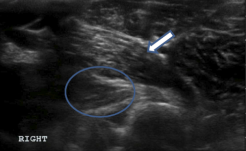
Longitudinal view of a right iliopsoas tendon of insertion. Note the hypoechoic changes of the fibrefibres consistent with a “core” lesion (circled). Lying overtop the iliopsoas tendon is the adductor muscle (white arrow).
CULLEN, Robert et al. Clinical Evaluation of Iliopsoas Strain with Findings from Diagnostic Musculoskeletal Ultrasound in Agility Performance Canines – 73 Cases. Veterinary Evidence, [S.l.], v. 2, n. 2, jun. 2017. ISSN 2396-9776.

Longitudinal (a) and cross-sectional (b) views of a Grade I right iliopsoas tendon injury. Note the mild hypoechoic changes but overall intact fibre pattern.
CULLEN, Robert et al. Clinical Evaluation of Iliopsoas Strain with Findings from Diagnostic Musculoskeletal Ultrasound in Agility Performance Canines – 73 Cases. Veterinary Evidence, [S.l.], v. 2, n. 2, jun. 2017. ISSN 2396-9776.

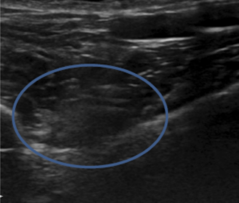
Oblique cross-sectional view of a Grade II left iliopsoas tendon injury. Note the hypoechoic changes and swelling with disruption of fascial lines (circled).
CULLEN, Robert et al. Clinical Evaluation of Iliopsoas Strain with Findings from Diagnostic Musculoskeletal Ultrasound in Agility Performance Canines – 73 Cases. Veterinary Evidence, [S.l.], v. 2, n. 2, jun. 2017. ISSN 2396-9776.

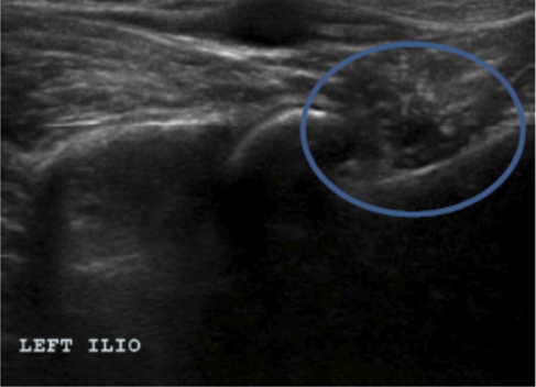
Longitudinal view of a Grade III left iliopsoas tendon injury. Note the hypoechoic changes and swelling with complete disruption of fascial lines (circled). This image shows a Grade III lesion not obtained from the case series presented in this paper, but is meant to illustrate the injury appearance.
CULLEN, Robert et al. Clinical Evaluation of Iliopsoas Strain with Findings from Diagnostic Musculoskeletal Ultrasound in Agility Performance Canines – 73 Cases. Veterinary Evidence, [S.l.], v. 2, n. 2, jun. 2017. ISSN 2396-9776.

Longitudinal views of the iliopsoas tendon of insertion of the left (labeled) and the normal right iliopsoas tendons of insertion. Note the hyperechoic changes of the fibres of the left tendon as well as irregularities of the lesser trochanter (white arrow).
CULLEN, Robert et al. Clinical Evaluation of Iliopsoas Strain with Findings from Diagnostic Musculoskeletal Ultrasound in Agility Performance Canines – 73 Cases. Veterinary Evidence, [S.l.], v. 2, n. 2, jun. 2017. ISSN 2396-9776.

A view of the iliacus and psoas major muscle bellies converging with an appreciation of the femoral nerve running between (circled). Note the thickened appearance of the nerve consistent with femoral neuritis and enlargement, an uncommon sequelae of iliopsoas inflammation.
CULLEN, Robert et al. Clinical Evaluation of Iliopsoas Strain with Findings from Diagnostic Musculoskeletal Ultrasound in Agility Performance Canines – 73 Cases. Veterinary Evidence, [S.l.], v. 2, n. 2, jun. 2017. ISSN 2396-9776.

Two longitudinal views of the left (labeled) and right iliopsoas tendons of insertion and bursas. Note the distended anechoic bursa on the right (white arrow) compared to the left.
CULLEN, Robert et al. Clinical Evaluation of Iliopsoas Strain with Findings from Diagnostic Musculoskeletal Ultrasound in Agility Performance Canines – 73 Cases. Veterinary Evidence, [S.l.], v. 2, n. 2, jun. 2017. ISSN 2396-9776.

Diese Webseite wurde mit Jimdo erstellt! Jetzt kostenlos registrieren auf https://de.jimdo.com


