Article added / Artikel hinzugefügt 01.10.2021
Generally Articles and Discussions about Osteosarcoma in Dogs
→ Evaluations of phylogenetic proximity in a group of 67 dogs with
osteosarcoma: a pilot study
Article added / Artikel hinzugefügt 01.10.2021
Generally Articles and Discussions about Osteosarcoma in Dogs
→ Canine Periosteal Osteosarcoma
Images added / Abbildungen hinzugefügt 02.05.2019
Generally Sonography Atlas of Dogs →
Cardiovascular system → Pulmonary vessels
New subcategory added / Neue Unterkategorie hinzugefügt 02.05.2019
Generally Sonography Atlas of Dogs →
Cardiovascular system → Pulmonary vessels
Images added / Abbildungen hinzugefügt 01.05.2019
Generally Sonography Atlas of Dogs →
Cardiovascular system → Heart valvular diseases


Generally Sonography Atlas of Dogs - Others
(Allgemeiner Sonographie-Atlas von Hunden - Sonstiges)
Thyroid
(Schilddrüse)
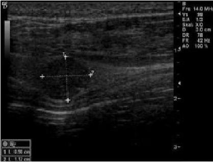
Ultraschallbild: Hund mit einem Tumor der Nebenschilddrüse
Mit Dank an die Tierklinik am Hasenberg in Stuttgart für die freundliche Unterstützung. Copyright (c) Tierklinik am Hasenberg, Stuttgart http://tierklinik-stuttgart.de

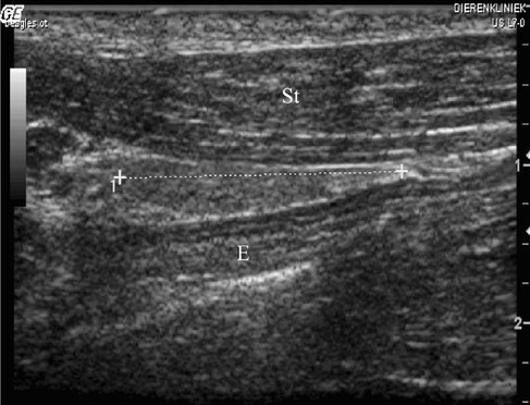
Longitudinal (left) and transverse
(right) ultrasound images of a normal left thyroid lobe obtained with a matrix line
ar transducer at 12 MHz. Cranial is left on the longitudinal image and medial is left on the transverse image. The linear scale on the right side of each image is in centimeters. The thyroid lobe
is indicated by electronic calipers. C, common carotid artery; E, esophagus; Sc, sternocephalic muscle; Sh,
sternohyoid muscle; St, sternothyroid muscle; T, trachea.
O. Taeymans, K. Peremans and J.H. Saunders
"Thyroid Imaging in the Dog: Current Status and Future Directions"
Article first published online: 28 JUN 2008
DOI: 10.1111/j.1939-1676.2007.tb03008.x

Longitudinal image of the left lobe (left side of the image) and transverse image of the right
lobe (right side of the image) in a primary hypothyroid Border Collie obtained with a matrix linear transdu
cer at 12 MHz. Both lobes are hypoechoic compared to the overlying sternothyroid muscles and have an inhomogeneous parenchyma. The gland has an irregular capsule on the
longitudinal image and
has a rounded shape on the transverse image. The size of both lobes was reduc
ed. C, common carotid artery; E, esophagus; St, sternothyroid muscle; T, trachea.
O. Taeymans, K. Peremans and J.H. Saunders
"Thyroid Imaging in the Dog: Current Status and Future Directions"
Article first published online: 28 JUN 2008
DOI: 10.1111/j.1939-1676.2007.tb03008.x

Long axis ultrasound image of the left thyroid lobe of a dog (small arrows). The ventral aspect of the neck is at the top of the image, and the cranial pole of the thyroid is to the left. A 3-4 mm hypoechoic mass is seen associated with the cranial pole of the thyroid (large arrow). Moderate farfield enhancement is also seem deep to the lesion.
Erik R. Wisner, "Clincal Vignette", Journal of Veterinary Internal Medicine, Vol8. No 3 (May-June), 1994: pp 244-245

(a) Longitudinal section of the right thyroid lobe in a euthyroid dog. The echotexture of the thyroid gland (*) was hyperechoic in
comparison with the adjacent sternothyroid muscle (1).
(b) Longitudinal section of the left thyroid lobe in a thyroglobulin autoantibody–
positive (TgAA-positive) hypothyroid dog. The echotexture of the thyroid gland (*) was hypoechoic in comparison with the adjacent
sternothyroid muscle (1)
(c) Longitudinal section of the left thyroid
gland (*) in a TgAA-negative hypothyroid dog. The echotexture of the thyroid gland was heterogeneous with
a hypoechoic background interrupted by hyperechoic lines and spots.
Reese, S., Breyer, U., Deeg, C., Kraft, W. and Kaspers, B. (2005), "Thyroid Sonography as an Effective Tool to Discriminate between Euthyroid Sick and Hypothyroid Dogs". Journal of Veterinary Internal Medicine, 19: 491–498. doi: 10.1111/j.1939-1676.2005.tb02717.x

Transversal plane of the left thyroid lobe in a euthyroid male beagle. The maximal cross sectional area (MCSA) of the thyroid gland (*) and of the sternothyroid muscle (1) were marked by dotted lines. 1, trachea; 2, esophagus; 3, left common carotid artery.
Reese, S., Breyer, U., Deeg, C., Kraft, W. and Kaspers, B. (2005), "Thyroid Sonography as an Effective Tool to Discriminate between Euthyroid Sick and Hypothyroid Dogs". Journal of Veterinary Internal Medicine, 19: 491–498. doi: 10.1111/j.1939-1676.2005.tb02717.x

Diese Webseite wurde mit Jimdo erstellt! Jetzt kostenlos registrieren auf https://de.jimdo.com


