

Article added / Artikel hinzugefügt 01.10.2021
Generally Articles and Discussions about Osteosarcoma in Dogs
→ Evaluations of phylogenetic proximity in a group of 67 dogs with
osteosarcoma: a pilot study
Article added / Artikel hinzugefügt 01.10.2021
Generally Articles and Discussions about Osteosarcoma in Dogs
→ Canine Periosteal Osteosarcoma
Images added / Abbildungen hinzugefügt 02.05.2019
Generally Sonography Atlas of Dogs →
Cardiovascular system → Pulmonary vessels
New subcategory added / Neue Unterkategorie hinzugefügt 02.05.2019
Generally Sonography Atlas of Dogs →
Cardiovascular system → Pulmonary vessels
Images added / Abbildungen hinzugefügt 01.05.2019
Generally Sonography Atlas of Dogs →
Cardiovascular system → Heart valvular diseases


Generally Sonography Atlas of Dogs - Others
(Allgemeiner Sonographie-Atlas von Hunden - Sonstiges)
Eye - Page 2
(Auge - Seite 2)

Photographic mage of left eye dog number 5. Note cataract focal immature (red arrow). ultrasound image in plan horizontal eye of the same dog. Note axial cortex and posterior pole crystalline hyperechoic (yellow arrow ) (cortical and polar cataract).
RODRIGUES JUNIOR, Emílio Fernandes. "Ultra-sonografia pré-cirúrgica da lente e do segmento posterior de cães portadores de catarata". 2008. xiii, 53 f. Dissertação (mestrado) - Universidade Estadual Paulista, Faculdade de Ciências Agrárias e Veterinárias, 2008.


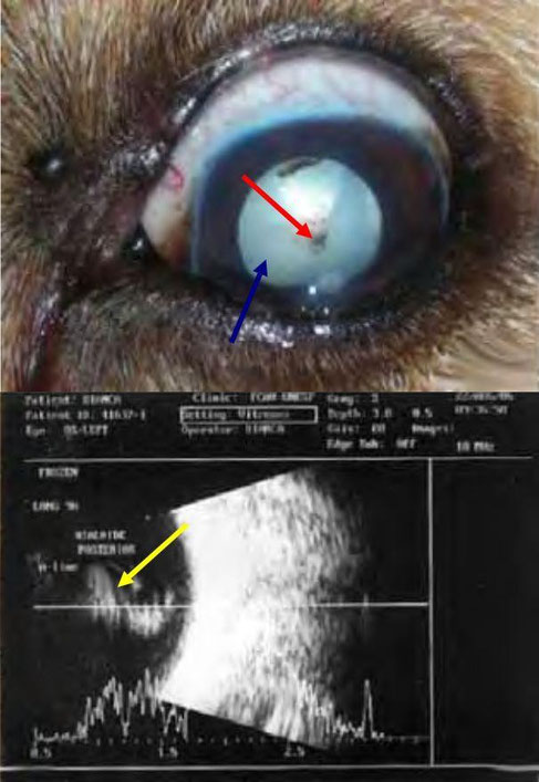
Photographic image of the left eye dog 9. Note mature cataract (red arrow) and conjunctival hyperemia (blue arrow). ultrasound image horizontal plane of the eye of the
same dog.
Notice preceding lenticular cortex
equatorial and axial hyperechoic (yellow arrow), posterior cortex and nucleus hypoechoic (green arrow) (complete cataract).
RODRIGUES JUNIOR, Emílio Fernandes. "Ultra-sonografia pré-cirúrgica da lente e do segmento posterior de cães portadores de catarata". 2008. xiii, 53 f. Dissertação (mestrado) - Universidade Estadual Paulista, Faculdade de Ciências Agrárias e Veterinárias, 2008.

Photographic mage of left eye dog number 1. Note pigment attached to the anterior lens capsule (red arrow) and mature cataract (blue arrow). ultrasound
image in the horizontal plane of the eye of the same dog.
Note the presence of persistent hyaloid artery attached to the capsule
lens and posterior papillary area (yellow arrow ).
RODRIGUES JUNIOR, Emílio Fernandes. "Ultra-sonografia pré-cirúrgica da lente e do segmento posterior de cães portadores de catarata". 2008. xiii, 53 f. Dissertação (mestrado) - Universidade Estadual Paulista, Faculdade de Ciências Agrárias e Veterinárias, 2008.


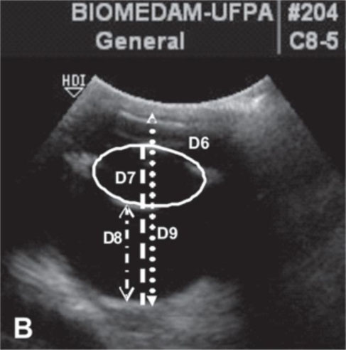
Measurements of the scheme of internal estruduras eyeball in canis familiaris adults.
A) D1 = thickness of the cornea ; D2 = the distance between the central point of the image of the cornea is the anterior lens capsule ; D3 = distance between the point of the image of the cornea and the posterior capsule of the lens ; D4 = lens thickness ; D5 = lens diameter
B ) D6 = area of the lens ; D7 = chamber glassy ; D8 = distance of the anterior lens capsule and retina ; D9 = distance between the image of the cornea and retina.
Beserra, Poliana S., Sales, Gustavo A., Santana, Expedito J.M., Miranda, Stefânia A., Brito, Adriel B., Nickolak, Elizabete, & Domingues, Sheyla F.S.. (2009). "Relação entre a biometria ultra-sonográfica em modo B do bulbo ocular e os diâmetros fronto occiptal e bizigomático em Canis familiaris." Pesquisa Veterinária Brasileira, 29(4), 286-290. Retrieved February 21, 2016, from http://www.scielo.br/scielo.php?script=sci_arttext&pid=S0100-736X2009000400002&lng=en&tlng=pt.


Ultrasound of the right eye of a Shih Tzu.
Ultrasound of the left eye of the same Shih Tzu.
With special thanks to Irene García Patiño (Sombra Acústica), veterinarian at the Veterinary Clinic Argos in Cee (A Coruña, Spain). http://sombraacustica.com


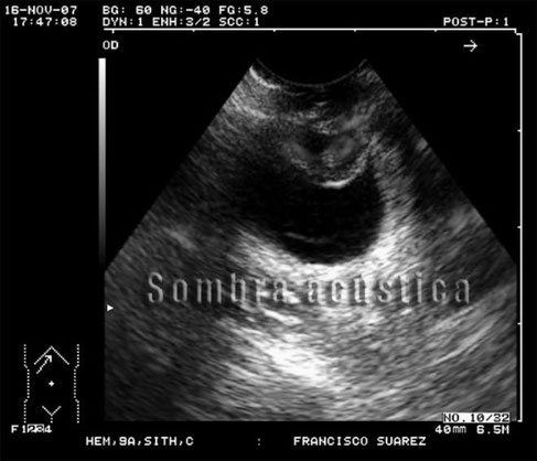
Cataract and lens luxation.
With special thanks to Irene García Patiño (Sombra Acústica), veterinarian at the Veterinary Clinic Argos in Cee (A Coruña, Spain). http://sombraacustica.com


Ultrasound image of a normal eye.
Mehdi Tavana and Seyedeh Zeinab Peighambarzadeh, "NORMAL OCULAR ULTRASONOGRAPHIC FINDING IN DOG", www.cibtech.org/sp.ed/jls/2014/03/jls.htm


Ultrasound image obtained with a 20 MHz transducer and the simultaneous use of A and B-modes showing an axial section of the right eye globe of a brachycephalic dog illustrating measurements obtained in the evaluation.
D1 corresponds to the distance between the cornea and
the anterior lens capsule; D2 the distance from the anterior capsule to the posterior lens capsule; D3 the distance from the posterior capsule to the posterior pole of the eye; and D4 the axial length of the
eye, from the
cornea to the posterior pole of the eye.
Meirelles, Adriana É. W. B., Toni, Maria C., Canola, Júlio C., Laus, José L., "Ophthalmic ultrasound of dogs with different skull conformations",Revista Brasileira de Ciências Agrárias [en linea] 2013, 8 ( ) : [Fecha de consulta: 6 de octubre de 2016] Disponible en:<http://www.redalyc.org/articulo.oa?id=119027922014> ISSN 1981-1160
