Article added / Artikel hinzugefügt 01.10.2021
Generally Articles and Discussions about Osteosarcoma in Dogs
→ Evaluations of phylogenetic proximity in a group of 67 dogs with
osteosarcoma: a pilot study
Article added / Artikel hinzugefügt 01.10.2021
Generally Articles and Discussions about Osteosarcoma in Dogs
→ Canine Periosteal Osteosarcoma
Images added / Abbildungen hinzugefügt 02.05.2019
Generally Sonography Atlas of Dogs →
Cardiovascular system → Pulmonary vessels
New subcategory added / Neue Unterkategorie hinzugefügt 02.05.2019
Generally Sonography Atlas of Dogs →
Cardiovascular system → Pulmonary vessels
Images added / Abbildungen hinzugefügt 01.05.2019
Generally Sonography Atlas of Dogs →
Cardiovascular system → Heart valvular diseases


Generally Sonography Atlas of Dogs - Genitourinary system
(Allgemeiner Sonographie-Atlas von Hunden) - (Urogenitales System)
Female reproduktion
(Weibliche Fortpflanzung)
Pyometra . U = cross sections of uterine horn .
N. Díez Bru
"Ecografía abdominal en pequeños animales."
CLINICA VETERINARIA DE PEQUEÑOS ANIMALES
Volumen 12, Número 3, Julio/Septiembre 1992

Ovarian cyst ( Q ) flow to the left kidney (R ).
N. Díez Bru
"Ecografía abdominal en pequeños animales."
CLINICA VETERINARIA DE PEQUEÑOS ANIMALES
Volumen 12, Número 3, Julio/Septiembre 1992

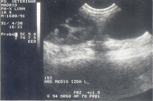
Mass ( M ) medial to the right kidney (R). Histopathological diagnosis : Cystadenocarcinoma of right ovary.
N. Díez Bru
"Ecografía abdominal en pequeños animales."
CLINICA VETERINARIA DE PEQUEÑOS ANIMALES
Volumen 12, Número 3, Julio/Septiembre 1992

Pyometra beim Hund. Mit U gekennzeichnet sind zwei Uterusschlingen, welche vom Schallkegel quer angeschnitten wurden.
http://de.wikipedia.org/wiki/Pyometra
„Pyometra Sono“. Lizenziert unter CC BY-SA 3.0 über Wikimedia Commons - http://commons.wikimedia.org/wiki/File:Pyometra_Sono.jpg#mediaviewer/File:Pyometra_Sono.jpg

Mehrere kleine Zysten im Eierstock
Mit Dank für die freundliche Genehmigung an die www.tierklinik.de

Eine grosse Zyste ausserhalb des Ovariums, eine kleinere innerhalb des Eierstocks.
Mit Dank für die freundliche Genehmigung an die www.tierklinik.de


Pathologische Veränderungen an der Gebärmutter. Hier in diesem Beispiel handelt es sich um eine Gebärmuttervereiterung, eine „Pyometra“ die man als eitergefüllte Hohlräume auf dem Bild schön erkennen kann.
Mit freundlicher Genehmigung der Tierarztpraxis Dr. Peter Neu, Coburg.

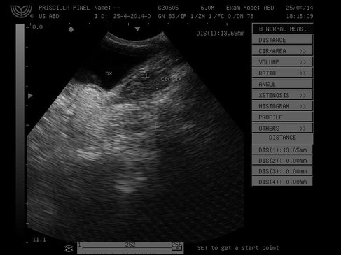
Image of a dog with content cellularity in the cervix after childbirth in clinical examination , it was observed mucopurulence ( provavelemente vaginitis ).
With special thanks to Priscilla Pinel, Medical Veterinary.
Currently serves in veterinary clinics and homes for the municipality of Rio de Janeiro ( south, north and west ).
http://veterinariapriscillapinel.com.br

Ovarian cancer.
With special thanks to Priscilla Pinel, Medical Veterinary.
Currently serves in veterinary clinics and homes for the municipality of Rio de Janeiro ( south, north and west ).
http://veterinariapriscillapinel.com.br

Ultra sonographic images of uterine involution in bitches after caesarean section , in transverse and longitudinal cuts, using a 7.5 MHz linear transducer.
A: Day 0, body. The luminal contents this relatively homogeneous and anechoic . The endometrium may be observed as hyperechoic ring surrounded by hypoechoic layer (myometrium ) , which in
turn , and surrounded by a hyperechoic line (serosa ), which notes the location of the suture.
B: Day 0, horn in longitudinal section , where it is observed the anechoic luminal contents and well-defined layers.
C: Day 3, horn
D: Day 3, near the bifurcation . The luminal contents this homogeneous surrounded by the endometrium and myometrium and this by serous.
E: Day 3 , bifurcation with observation of the suture in the ventral portion.
Ferri, S.T.S., Vicente, W.R.R., & Toniollo, G.H.. (2003). "Estudo da involução uterina por meio da ultra-sonografia (modo-B) em cadelas submetidas a cesariana". Arquivo Brasileiro de Medicina Veterinária e Zootecnia, 55(2), 167-172. Retrieved January 23, 2016, from http://www.scielo.br/scielo.php?script=sci_arttext&pid=S0102-09352003000200007&lng=en&tlng=pt.

Uterine involution of ultrasound images of bitches after caesarean section , in cross-sections , using linear and sector transducer of 7.5 MHz .
A: Day 7 , uterine horn , with sector transducer .
B: Day 7 , near the bifurcation .
C: Day 14 , horn.
D: Day 14 , horn.
E: Day 21 horn. The luminal contents this relatively homogeneous and hyperechoic , mingling with the endometrium . Uterine layers station thin and lumen colapsado.O hyperechoic ring surrounding the myometrium not that well defined.
Ferri, S.T.S., Vicente, W.R.R., & Toniollo, G.H.. (2003). "Estudo da involução uterina por meio da ultra-sonografia (modo-B) em cadelas submetidas a cesariana". Arquivo Brasileiro de Medicina Veterinária e Zootecnia, 55(2), 167-172. Retrieved January 23, 2016, from http://www.scielo.br/scielo.php?script=sci_arttext&pid=S0102-09352003000200007&lng=en&tlng=pt.

Ultrasound images (ecovet Canarias ) of three female dogs with pyometra.
With special thanks to Irene García Patiño (Sombra Acústica), veterinarian at the Veterinary Clinic Argos in Cee (A Coruña, Spain). http://sombraacustica.com

Female Poodle 18 years with clinical suspicion of closed pyometra (uterine infection).
In clinical examination showed abdominal pain and seizure.
In sonographic examination was observed increase in the uterus and hyperechoic walls and
thickened with moderate abdominal free liquid and still had a heterogeneous mass with
irregular borders. Apart from gastroenteritis and chronic kidney disease.
With special thanks to Priscilla Pinel, Medical Veterinary.
Currently serves in veterinary clinics and homes for the municipality of Rio de Janeiro ( south, north and west ).
http://veterinariapriscillapinel.com.br


A sonographic view of uterus of Griffon dog 10-years old showing: Hypoechoic and thickened uterine wall. The luminal content was homogenous and filled with anechoic fluid.
F.Tawfik M, Oda SS, El-Neweshy MS, El-Manakhly EM. " Pathological Study on Female Reproductive Affections in Dogs and Cats at Alexandria Province", Egypt. www.scopemed.org/?mno=187841 [Access: September 21, 2016]. doi:10.5455/ajvs.187841

Diese Webseite wurde mit Jimdo erstellt! Jetzt kostenlos registrieren auf https://de.jimdo.com


