Article added / Artikel hinzugefügt 01.10.2021
Generally Articles and Discussions about Osteosarcoma in Dogs
→ Evaluations of phylogenetic proximity in a group of 67 dogs with
osteosarcoma: a pilot study
Article added / Artikel hinzugefügt 01.10.2021
Generally Articles and Discussions about Osteosarcoma in Dogs
→ Canine Periosteal Osteosarcoma
Images added / Abbildungen hinzugefügt 02.05.2019
Generally Sonography Atlas of Dogs →
Cardiovascular system → Pulmonary vessels
New subcategory added / Neue Unterkategorie hinzugefügt 02.05.2019
Generally Sonography Atlas of Dogs →
Cardiovascular system → Pulmonary vessels
Images added / Abbildungen hinzugefügt 01.05.2019
Generally Sonography Atlas of Dogs →
Cardiovascular system → Heart valvular diseases


Generally Sonography Atlas of Dogs - Abdomen
(Allgemeiner Sonographie-Atlas von Hunden) - (Abdomen)
Liver - Page 2
(Leber - Seite 2)
Liver tumors (rounded nodules).
With special thanks to Priscilla Pinel, Medical Veterinary.
Currently serves in veterinary clinics and homes for the municipality of Rio de Janeiro ( south, north and west ).
http://veterinariapriscillapinel.com.br

Ultrasound images of dog liver .
A: left portal branch (RPE) parallel to the left hepatic vein (HEV)
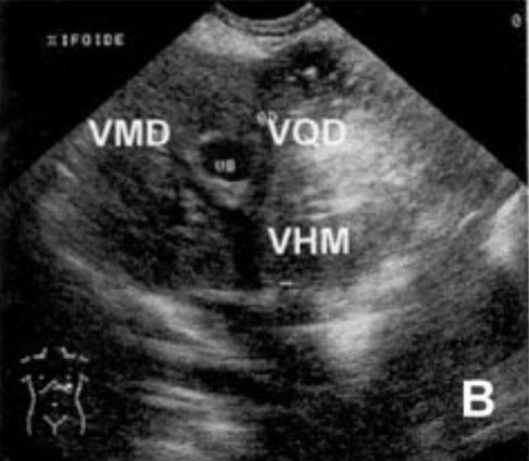
B: middle hepatic vein (WHV) formed by
confluence of the hepatic vein of the right medial lobe (VMD) and hepatic vein Square wolf (VQD)
C: vena cava flow (VCC) , hepatic vein side lobe (VLD) hepatic vein and the caudate process ( VCD)
D : obvious portal vessels (VP)
E: no obvious portal vessels (VP)
F : vein foramen cava flow in the diaphragm (Fo) .
Annelise Baldacin Salgado,Patrícia Reginato Facciotti,Dulcinéa Gon?alves Teixeira,Karla Patrícia Cardoso Araújo
"IDENTIFICA O DAS REGI ES CORRESPON-DENTES AOS LOBOS HEPáTICOS DE C ES POR MEIO DA ULTRA-SONOGRAFIA"
Ciência Animal Brasileira, v. 8, n. 3, p. 545-558, jul./set. 2007

Positioning the transducer and
respective ultrasound images.
A: left intercostal window, the liver was observed (F) and stomach (E)
B: subcostal parasternal left liver was observed (F) and stomach (E)
C: paravertebral right flow window , it is observed liver (F) , right kidney (RD) and caudal vena cava (VCC)
D: paravertebral right cranial window , the liver was observed (F) , lung (Pu) and vena cava flow rate (VCC)
E: right parasternal window (side), it is observed liver (F) with gallbladder
(VB) to the left
F: right parasternal window (medial) is observed liver (F) with gall bladder (VB) to the right
Annelise Baldacin Salgado,Patrícia Reginato Facciotti,Dulcinéa Gon?alves Teixeira,Karla Patrícia Cardoso Araújo
"IDENTIFICA O DAS REGI ES CORRESPON-DENTES AOS LOBOS HEPáTICOS DE C ES POR MEIO DA ULTRA-SONOGRAFIA"
Ciência Animal Brasileira, v. 8, n. 3, p. 545-558, jul./set. 2007

1. ventrocranial right segment - medial lobe with its left edge in contact with the gallbladder (Fig. B), corresponds to the intercostal
parasternal right window (side) 2. ventrocaudal right segment - medial lobe flow to the gallbladder in his caudal- dorsal portion is the pyloric region and duodenum (Fig. B),
corresponds to the region flow and intercostal parasternal right window (side)
3. Left ventrocranial segment - left medial lobe (Fig. B), corresponds to the cranial region, and the left intercostal window 4. Left ventrocaudal segment
- left lateral lobe, in his caudal- dorsal portion. They are the stomach and caudo-ventral the spleen (Fig. B), corresponds to the intercostal window left, which, according to our results,
identifies the diaphragmatic face of this wolf, and the subcostal left parasternal window, which identifies the visceral surface 5. ventrocranial segment through - Wolf
square most of the segment, but cranial to the gallbladder , medial lobe, gallbladder in contact with the right side edge of the square lobe (Fig. B), corresponds to the intercostal parasternal
right (medial) 6. ventrocaudal middle segment - predominantly left lateral lobe, dorsally is the body of the stomach (Fig. B), corresponds to the caudal region to the intercostal
window parasternal right (medial) 7. right dorsocranial segment - medial lobe right cranial to the neck of the gallbladder (Fig. C)
and corresponds to the cranial region of paravertebral right cranial window 8. right dorso-caudal segment - lateral lobe left and caudate process of the caudate lobe, the left
lateral lobe occupies the region more cranial and lateral caudate process occupies the caudal and medial region with its renal impression in contact with the cranial end of the right kidney;
ventral is the pylorus and duodenum region (Fig. C). This segment corresponds to two windows, the paraspinal cranial right, which, as the results of this work relates to the side right back, and
paraspinal right flow, where it was not possible to differentiate between the lateral lobe and caudate process 9. left dorsocranial segment - left medial lobe in the most cranial
portion and the left side at the flow rate; in your area flow has a large contact area with the stomach (Fig. C), corresponds to the region craniodorsal left intercostal window
10. the left dorso-caudal segment - process papillary the caudate lobe in cranio-medial region; the rest of the body and thread are the fundus (Fig. C), stands for dorsal left
intercostal window 11. segment dorsocranial through - Wolf right medial (Fig. C); stands for dorsal to the intercostal parasternal right (medial) 12. dorso-caudal segment
through - body gastric (Fig. C); corresponds to the region dorsal-caudal to the intercostal parasternal right (medial)
Annelise Baldacin Salgado,Patrícia Reginato Facciotti,Dulcinéa Gon?alves Teixeira,Karla Patrícia Cardoso Araújo
"IDENTIFICA O DAS REGI ES CORRESPON-DENTES AOS LOBOS HEPáTICOS DE C ES POR MEIO DA ULTRA-SONOGRAFIA"
Ciência Animal Brasileira, v. 8, n. 3, p. 545-558, jul./set. 2007

Hepatomegaly. Irregular liver texture, ascites.
2 days after the first ultrasound.
With special thanks to Irene García Patiño (Sombra Acústica), veterinarian at the Veterinary Clinic Argos in Cee (A Coruña, Spain). http://sombraacustica.com

Congenital Extrahepatic Shunt in a Puppy.
With special thanks to the authors of the book "Small Animal Ultrasonography" , Marc-André d’Anjou and Dominique Penninck

Transverse sonogram of the liver demon-strating a round lesion (straight arrows) containing mixed echoes. Note the highly echogenic interface (curved arrow) with distal shadowing at the nondependent margin, indicating gas within the lesion.
Grooters, A. M., Sherding, R. G., Biller, D. S. and Johnson, S. E. (1994), "Hepatic Abscesses Associated With Diabetes Mellitus in Two Dogs". Journal of Veterinary Internal Medicine, 8: 203–206. doi: 10.1111/j.1939-1676.1994.tb03216.x

Longitudinal sooogram of the liver (case 2) showing a hypoechoic lesion in the liver.
Grooters, A. M., Sherding, R. G., Biller, D. S. and Johnson, S. E. (1994), "Hepatic Abscesses Associated With Diabetes Mellitus in Two Dogs". Journal of Veterinary Internal Medicine, 8: 203–206. doi: 10.1111/j.1939-1676.1994.tb03216.x

A conventional US liver image with 3 hypoechoic lesions. Two small (asterisk) lesions and one large (white arrows)
targeted lesion.
Wdowiak, M., Rychlik, A., Nieradka, R., et al. (2010). "Contrast-enhanced ultrasonography (CEUS) in canine liver examination". doi:10.2478/v10181-010-0007-2

Late phase CEUS image. The isoecho- genic liver parenchyma indicates the benign nature of the hypoechoic lesions.
Wdowiak, M., Rychlik, A., Nieradka, R., et al. (2010). "Contrast-enhanced ultrasonography (CEUS) in canine liver examination". doi:10.2478/v10181-010-0007-2

Ultrasonograph (2D) in sagittal scan of shrunk liver with hyperechoic hepatic parenchyma and irregular lobe margins surrounded with anechoic abdominal effusion in a 5-year-old intact male Cocker spaniel dog.
Vijay Kumar, Adarsh Kumar, A. C. Varshney, S. P. Tyagi, M. S. Kanwar, and S. K. Sharma, “Diagnostic Imaging of Canine Hepatobiliary Affections: A Review,” Veterinary Medicine International, vol. 2012, Article ID 672107, 15 pages, 2012. doi:10.1155/2012/672107

Sonograph (2D) in transverse scan showing hyperechoic liver lobe and cholecystitis with inspissated bile in gall bladder (GB) in a 7-year-old intact Pomeranian male dog.
Vijay Kumar, Adarsh Kumar, A. C. Varshney, S. P. Tyagi, M. S. Kanwar, and S. K. Sharma, “Diagnostic Imaging of Canine Hepatobiliary Affections: A Review,” Veterinary Medicine International, vol. 2012, Article ID 672107, 15 pages, 2012. doi:10.1155/2012/672107

Dorsal sonogram (2D) of liver depicting hypoechoic parenchyma with portal vessel dilatation in one-year-old mixed-breed dog.
Vijay Kumar, Adarsh Kumar, A. C. Varshney, S. P. Tyagi, M. S. Kanwar, and S. K. Sharma, “Diagnostic Imaging of Canine Hepatobiliary Affections: A Review,” Veterinary Medicine International, vol. 2012, Article ID 672107, 15 pages, 2012. doi:10.1155/2012/672107

Longitudinal sonogram (2D) of liver depicting hypoechoic parenchyma with marked portal vessel dilatation (subjective) in 5-year-old mixed-breed dog with significant spleenomegaly.
Vijay Kumar, Adarsh Kumar, A. C. Varshney, S. P. Tyagi, M. S. Kanwar, and S. K. Sharma, “Diagnostic Imaging of Canine Hepatobiliary Affections: A Review,” Veterinary Medicine International, vol. 2012, Article ID 672107, 15 pages, 2012. doi:10.1155/2012/672107

Transverse sonogram (2D) of right medial liver lobe of two and half- year-old intact female Pointer dog depicting generalized hyperechoic parenchyma with biliary sludge in gall bladder.
Vijay Kumar, Adarsh Kumar, A. C. Varshney, S. P. Tyagi, M. S. Kanwar, and S. K. Sharma, “Diagnostic Imaging of Canine Hepatobiliary Affections: A Review,” Veterinary Medicine International, vol. 2012, Article ID 672107, 15 pages, 2012. doi:10.1155/2012/672107

Two-dimensional ultrasonographic appearance of liver in sagittal scan depicting hyperechoic parenchyma with rounding of liver lobe surrounded with textured fluid in a 6-year-old male Labrador Retriever affected with infectious peritonitis.
Vijay Kumar, Adarsh Kumar, A. C. Varshney, S. P. Tyagi, M. S. Kanwar, and S. K. Sharma, “Diagnostic Imaging of Canine Hepatobiliary Affections: A Review,” Veterinary Medicine International, vol. 2012, Article ID 672107, 15 pages, 2012. doi:10.1155/2012/672107

Hepatic sonogram (2D) in sagittal scan of a five-year-old intact Pomeranian male dog depicting increased parenchymal echogenicity with hyperechoic double rim appearance of GB wall.
Vijay Kumar, Adarsh Kumar, A. C. Varshney, S. P. Tyagi, M. S. Kanwar, and S. K. Sharma, “Diagnostic Imaging of Canine Hepatobiliary Affections: A Review,” Veterinary Medicine International, vol. 2012, Article ID 672107, 15 pages, 2012. doi:10.1155/2012/672107

Two-dimensional ultrasonographic appearance in sagittal scan of cirrhosed liver lobe in a seven-year-old intact mixed-breed dog depicting collapsed and irregularly shaped gall bladder with echogenic and thickened (4 mm) GB wall.
Vijay Kumar, Adarsh Kumar, A. C. Varshney, S. P. Tyagi, M. S. Kanwar, and S. K. Sharma, “Diagnostic Imaging of Canine Hepatobiliary Affections: A Review,” Veterinary Medicine International, vol. 2012, Article ID 672107, 15 pages, 2012. doi:10.1155/2012/672107

Longitudinal sonogram (2D) of liver of two and half-year-old, intact female Pointer dog showing cholecystitis as echogenic thickened GB wall and mineralization in the neck of gall bladder with acoustic shadowing along with hyperechoic parenchyma.
Vijay Kumar, Adarsh Kumar, A. C. Varshney, S. P. Tyagi, M. S. Kanwar, and S. K. Sharma, “Diagnostic Imaging of Canine Hepatobiliary Affections: A Review,” Veterinary Medicine International, vol. 2012, Article ID 672107, 15 pages, 2012. doi:10.1155/2012/672107

Transverse ultrasonogram (2D) of liver in a nine-year-old spayed female, German shepherd dog revealing two hyperechoic homogenous circular nodular masses measuring 55 mm and 29 mm.
Vijay Kumar, Adarsh Kumar, A. C. Varshney, S. P. Tyagi, M. S. Kanwar, and S. K. Sharma, “Diagnostic Imaging of Canine Hepatobiliary Affections: A Review,” Veterinary Medicine International, vol. 2012, Article ID 672107, 15 pages, 2012. doi:10.1155/2012/672107

Sagittal sonogram (2D) of liver in a ten-year-old intact Labrador retriever dog affected by a perianal tumour showing hyperechoic hepatic mass with central lytic changes.
Vijay Kumar, Adarsh Kumar, A. C. Varshney, S. P. Tyagi, M. S. Kanwar, and S. K. Sharma, “Diagnostic Imaging of Canine Hepatobiliary Affections: A Review,” Veterinary Medicine International, vol. 2012, Article ID 672107, 15 pages, 2012. doi:10.1155/2012/672107

Two-dimensional ultrasonographic transverse scan in a five-year-old intact mixed-breed dog showing an encap-sulated hypoechoic hepatic mass with irregular margins.
Vijay Kumar, Adarsh Kumar, A. C. Varshney, S. P. Tyagi, M. S. Kanwar, and S. K. Sharma, “Diagnostic Imaging of Canine Hepatobiliary Affections: A Review,” Veterinary Medicine International, vol. 2012, Article ID 672107, 15 pages, 2012. doi:10.1155/2012/672107

Sagittal sonograph (2D) of liver with extensive noncystic cavitary lesions in hepatic parenchyma in a nine-year-old intact male Boxer dog.
Vijay Kumar, Adarsh Kumar, A. C. Varshney, S. P. Tyagi, M. S. Kanwar, and S. K. Sharma, “Diagnostic Imaging of Canine Hepatobiliary Affections: A Review,” Veterinary Medicine International, vol. 2012, Article ID 672107, 15 pages, 2012. doi:10.1155/2012/672107

Two-dimensional ultrasonographic sagittal scan of right liver lobe in seven-year-old mixed-breed intact male dog showing noncystic cavitary lesions with generalized increase in paren-chymal echogenicity.
Vijay Kumar, Adarsh Kumar, A. C. Varshney, S. P. Tyagi, M. S. Kanwar, and S. K. Sharma, “Diagnostic Imaging of Canine Hepatobiliary Affections: A Review,” Veterinary Medicine International, vol. 2012, Article ID 672107, 15 pages, 2012. doi:10.1155/2012/672107

Ultrasonogram (2D) of liver in longitudinal scan revealing large poorly echogenic area with internal septations in a one and half-year-old neutered male Doberman pinscher dog.
Vijay Kumar, Adarsh Kumar, A. C. Varshney, S. P. Tyagi, M. S. Kanwar, and S. K. Sharma, “Diagnostic Imaging of Canine Hepatobiliary Affections: A Review,” Veterinary Medicine International, vol. 2012, Article ID 672107, 15 pages, 2012. doi:10.1155/2012/672107

Ultrasonographic image showing anechoeic fluid with floating liver.
Kumar KS, Srikala D. "Ascites with right heart failure in a dog:diagnosis and management". www.scopemed.org/?mno=157163 [Access: September 23, 2016]. doi:10.5455/javar.2014.a15

Transverse ultrasonographic view of the right liver of the dog, showing an extremely heterogeneous and irregularly marginated liver.
Zaheda Khan, Kathryn Gates, Stephen A Simpson: "Bicavitary effusion secondary to liver lobe torsion in a dog"
https://dx.doi.org/10.2147/VMRR.S83608

Ultrasonograph (2D) in sagittal scan of shrunk liver with hyperechoic hepatic parenchyma and irregular lobe margins surrounded with anechoic abdominal effusion in a 5-year-old intact male Cocker spaniel dog.
Kumar V, Kumar A, Varshney AC, Tyagi SP, Kanwar MS, Sharma SK. Diagnostic Imaging of Canine Hepatobiliary Affections: A Review. Veterinary Medicine International. 2012;2012:672107. doi:10.1155/2012/672107.

Transverse ultrasonogram (2D) of liver in a nine-year-old spayed female, German shepherd dog revealing two hyperechoic homogenous circular nodular masses measuring 55 mm and 29 mm.
Kumar V, Kumar A, Varshney AC, Tyagi SP, Kanwar MS, Sharma SK. Diagnostic Imaging of Canine Hepatobiliary Affections: A Review. Veterinary Medicine International. 2012;2012:672107. doi:10.1155/2012/672107.

Sagittal sonogram (2D) of liver in a ten-year-old intact Labrador retriever dog affected by a perianal tumour showing hyperechoic hepatic mass with central lytic changes.
Kumar V, Kumar A, Varshney AC, Tyagi SP, Kanwar MS, Sharma SK. Diagnostic Imaging of Canine Hepatobiliary Affections: A Review. Veterinary Medicine International. 2012;2012:672107. doi:10.1155/2012/672107.

Two-dimensional ultrasonographic transverse scan in a five-year-old intact mixed-breed dog showing an encapsu- lated hypoechoic hepatic mass with irregular margins.
Kumar V, Kumar A, Varshney AC, Tyagi SP, Kanwar MS, Sharma SK. Diagnostic Imaging of Canine Hepatobiliary Affections: A Review. Veterinary Medicine International. 2012;2012:672107. doi:10.1155/2012/672107.

Sagittal sonograph (2D) of liver with extensive noncystic cavitary lesions in hepatic parenchyma in a nine-year-old intact male Boxer dog.
Kumar V, Kumar A, Varshney AC, Tyagi SP, Kanwar MS, Sharma SK. Diagnostic Imaging of Canine Hepatobiliary Affections: A Review. Veterinary Medicine International. 2012;2012:672107. doi:10.1155/2012/672107.

Two-dimensional ultrasonographic sagittal scan of right liver lobe in seven-year-old mixed-breed intact male dog showing noncystic cavitary lesions with generalized increase in paren-chymal echogenicity.
Kumar V, Kumar A, Varshney AC, Tyagi SP, Kanwar MS, Sharma SK. Diagnostic Imaging of Canine Hepatobiliary Affections: A Review. Veterinary Medicine International. 2012;2012:672107. doi:10.1155/2012/672107.

Diese Webseite wurde mit Jimdo erstellt! Jetzt kostenlos registrieren auf https://de.jimdo.com


