

Article added / Artikel hinzugefügt 01.10.2021
Generally Articles and Discussions about Osteosarcoma in Dogs
→ Evaluations of phylogenetic proximity in a group of 67 dogs with
osteosarcoma: a pilot study
Article added / Artikel hinzugefügt 01.10.2021
Generally Articles and Discussions about Osteosarcoma in Dogs
→ Canine Periosteal Osteosarcoma
Images added / Abbildungen hinzugefügt 02.05.2019
Generally Sonography Atlas of Dogs →
Cardiovascular system → Pulmonary vessels
New subcategory added / Neue Unterkategorie hinzugefügt 02.05.2019
Generally Sonography Atlas of Dogs →
Cardiovascular system → Pulmonary vessels
Images added / Abbildungen hinzugefügt 01.05.2019
Generally Sonography Atlas of Dogs →
Cardiovascular system → Heart valvular diseases


Generally Sonography Atlas of Dogs - Abdomen
(Allgemeiner Sonographie-Atlas von Hunden) - (Abdomen)
Alimentary tract
(Verdauungstrakt)

Esophageal mass in a 14-year-old Welsh Corgi. A and B: Thoracic radiographs reveal a homogeneous, soft-tissue opacity (M, arrows) associated with the caudal
mediastinum, in the caudal region of the esophagus (E), silhouetting with the diaphragm. The position of the ultrasound transducer is shown in B. C and D:
Ultrasonographic and enhanced labeled images obtained with a ventral transhepatic approach (sagittal plane). At the level of the esophageal hiatus, the mass (M, arrows) is associated with the
esophagus (E). Liver and stomach (S) are visible in the near field, and the lung-diaphragm interface (D) manifests as a curvilinear, strongly hyperechoic structure bordering the esophageal
mass.
With special thanks to the authors of the book "Small Animal Ultrasonography" , Marc-André d’Anjou and Dominique Penninck


Darmverschluss durch einen Fremdkörper
(veröffentlicht mit freundlicher Genehmigung der Kleintierpraxis Schaub, Jena, http://www.tierarztpraxis-schaub.de)


Mucosal hyperechoic striations. Longitudinal (A) and transverse (B) sonograms of thickened jejunal segments with hyperechoic linear striations within the mucosal layer of a 5 year old French Bulldog. The striations represent dilated lacteals. This dog had a long history of inflammatory bowel disease. Peritoneal effusion is present (*). C and D: Similar but more severe findings are present in this 9 year old Jack Russell terrier presented with protein losing enteropathy. Note the thickened and hyperechoic adjacent fat (F), often present in these cases.
With special thanks to the authors of the book "Small Animal Ultrasonography" , Marc-André d’Anjou and Dominique Penninck


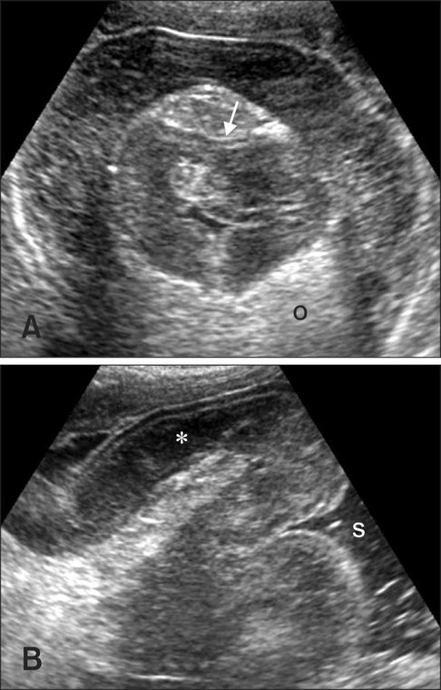
Ultrasonography of a pylorogastric intussusception in case 1. (A) A mass (m), 38 × 31 mm in size, connected with the descending duodenum (d). The mass occupied the body of the stomach (s). (B) Concentric multiple rings (arrow heads) represent an intussusception. The pyloric wall (double-headed arrow and W) is thickened (7.1 mm) with indistinct layering. Short arrows are multiple hypoechoic regions in the pyloric wall. Note the edematous change of mesentery (o).
Jihye Choi, Seoyeon Keh, Taeeun Kim, Jaeyoung Jang, Hyunwook Kim, Junghee Yoon
15 June 2012
3386350
Journal of Veterinary Science
The Korean Society of Veterinary Science

Ultrasonography a pylorogastric intussusception in case 2. (A) In the transverse view, a concentric, multiple ring (arrow) and edematous mesentery (o) was observed in the gastric body. (B) In longitudinal image of the ring, the invaginated pylorus showed thickened muscular layer (*) with distinct layering. The stomach (s) was dilated with fluid.
Jihye Choi, Seoyeon Keh, Taeeun Kim, Jaeyoung Jang, Hyunwook Kim, Junghee Yoon
15 June 2012
3386350
Journal of Veterinary Science
The Korean Society of Veterinary Science


Intestinal vascular anomaly in a 9 month old Labrador dog with vomiting and diarrhea.
A and B: Transverse sonograms of one fluid filled colonic segment with a thickened, nodular-like wall made of intramural enlarged varices (arrows in A), as confirmed using color flow Doppler (in B). A large network of numerous anomalous small vessels was seen throughout the abdomen, especially in the mesentery and near the main portal vein. C: Transverse post-contrast CT image of the caudal abdomen showing the intramural colonic vessels (arrow). D: Reconstructed CT dorsal image illustrating the numerous anomalous enlarged and tortuous vessels supportive of portal hypertension and acquired portosystemic shunts. Note the intramural colonic vessels (arrows). The left side of the liver is nearly absent. Because of the guarded prognosis, the dog was euthanatized and the final diagnosis is arteriovenous malformation with marked arteriolar proliferation and hypertrophy, venous ectasia, lobular atrophy and fibrosis.
With special thanks to the authors of the book "Small Animal Ultrasonography" , Marc-André d’Anjou and Dominique Penninck


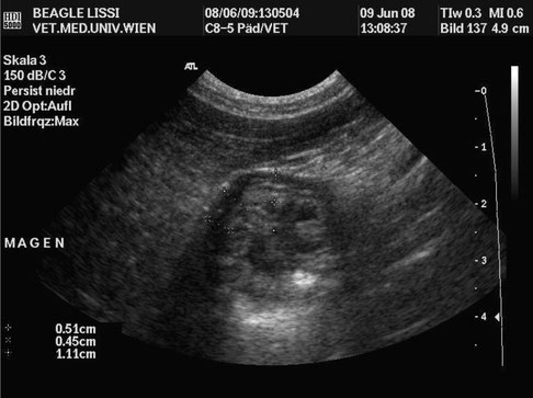
Ultraschallbild des Magens mit Gas im Lumen, transversal
Nödl, Barbara: Sonographische Untersuchungen zum Einfluss von DDGS auf den Magen-Darm-Trakt von Hunden.
Diplomarbeit, Vet. Med. Univ. Wien, pp. 41.

Ultraschallbild des Pylorus, transversal
Nödl, Barbara: Sonographische Untersuchungen zum Einfluss von DDGS auf den Magen-Darm-Trakt von Hunden.
Diplomarbeit, Vet. Med. Univ. Wien, pp. 41.


Ultraschallbild des Duodenums, sagittal, mit Messung der Wand
Nödl, Barbara: Sonographische Untersuchungen zum Einfluss von DDGS auf den Magen-Darm-Trakt von Hunden.
Diplomarbeit, Vet. Med. Univ. Wien, pp. 41.


Ultraschallbild des Dünndarms, sagittal, mit Messung der Wand
Nödl, Barbara: Sonographische Untersuchungen zum Einfluss von DDGS auf den Magen-Darm-Trakt von Hunden.
Diplomarbeit, Vet. Med. Univ. Wien, pp. 41.


Ultraschallbild des Magens, transversal, mit Messung der Wand
Nödl, Barbara: Sonographische Untersuchungen zum Einfluss von DDGS auf den Magen-Darm-Trakt von Hunden.
Diplomarbeit, Vet. Med. Univ. Wien, pp. 41.


Ultrasonographic images in the sagittal orientation of the umbilical region (A) and left lumbar (B and C) showing free abdominal air (arrows) that led to increased echogenicity
and thickening of the peritoneal stripe with reverberation artifacts. Free abdominal air was present along the ventral peritoneum adjacent to the spleen and small intestine. Intraluminal air was
also observed in the intestinal lumen (arrowhead) and associated with “dirty” acoustic shadowing.
Song Yeon Kim, Ki Tae Park, Seong Chan Yeon, Hee Chun Lee, "Accuracy of
sonographic diagnosis of pneumoperitoneum using the enhanced peritoneal stripe sign in beagle dogs", https://www.scienceopen.com/document/vid/d6821c71-e9b0-4baf-93d0-4aa4fe5cbeeb#main-article-text


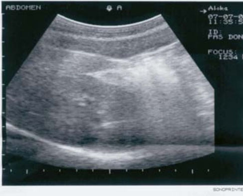
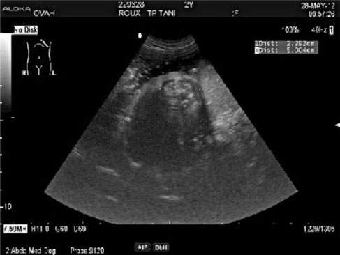
Physiological stomach
Krstić Vanja
"ENDOSCOPIC AND ULTRASOUND DIAGNOSTICS AS CONTEMPORARY
METHOD IN DIAGNOSTICS OF DOG STOMACH DISEASES", http://dx.doi.org/10.2298/VETGL0502141K

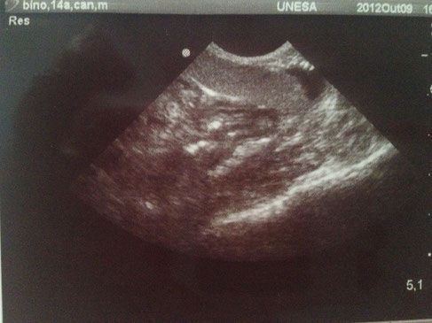
Dog stomach without water
Krstić Vanja
"ENDOSCOPIC AND ULTRASOUND DIAGNOSTICS AS CONTEMPORARY
METHOD IN DIAGNOSTICS OF DOG STOMACH DISEASES", http://dx.doi.org/10.2298/VETGL0502141K


Dog stomach filled with water
Krstić Vanja
"ENDOSCOPIC AND ULTRASOUND DIAGNOSTICS AS CONTEMPORARY
METHOD IN DIAGNOSTICS OF DOG STOMACH DISEASES", http://dx.doi.org/10.2298/VETGL0502141K


Ultrasound finding of ulcer in stomach
wall and coagulum
V. Krstic, N. Krstic, V. Ilic, Milena Dordevic
"RADIOLOGICAL, ULTRASOUND AND ENDOSCOPIC DIAGNOSTICS OF
CERTAIN CANINE STOMACH DISEASES"
UDK 636.7.09:616.33-07


Stomachs of three dogs with gastritis at different stages
With special thanks to Priscilla Pinel, Medical Veterinary.
Currently serves in veterinary clinics and homes for the municipality of Rio de Janeiro ( south, north and west ).
http://veterinariapriscillapinel.com.br


Images of colitis in dogs.
With special thanks to Priscilla Pinel, Medical Veterinary.
Currently serves in veterinary clinics and homes for the municipality of Rio de Janeiro ( south, north and west ).
http://veterinariapriscillapinel.com.br


Stomach wall of a dog thickened with large amounts of liquid and gas inside.
With special thanks to Priscilla Pinel, Medical Veterinary.
Currently serves in veterinary clinics and homes for the municipality of Rio de Janeiro ( south, north and west ).
http://veterinariapriscillapinel.com.br


Bowel loops with thickened wall and liquid content in the lumen of the same dog.
With special thanks to Priscilla Pinel, Medical Veterinary.
Currently serves in veterinary clinics and homes for the municipality of Rio de Janeiro ( south, north and west ).
http://veterinariapriscillapinel.com.br

Intestinal handles with pleated net free in the cavity (possible foreign body).
With special thanks to Priscilla Pinel, Medical Veterinary.
Currently serves in veterinary clinics and homes for the municipality of Rio de Janeiro ( south, north and west ).
http://veterinariapriscillapinel.com.br


Free liquid with red blood cell content in the abdomen (hemoabdomen).
With special thanks to Priscilla Pinel, Medical Veterinary.
Currently serves in veterinary clinics and homes for the municipality of Rio de Janeiro ( south, north and west ).
http://veterinariapriscillapinel.com.br


Notable intestinal dilatation, possible presence of a foreign body in the intestine.
With special thanks to Irene García Patiño (Sombra Acústica), veterinarian at the Veterinary Clinic Argos in Cee (A Coruña, Spain). http://sombraacustica.com


Dog, 3 yrs., with abdominal pain, vomiting and diarrhea for several days.
Sonographic examination shows an intestinal intussusception.
With special thanks to Irene García Patiño (Sombra Acústica), veterinarian at the Veterinary Clinic Argos in Cee (A Coruña, Spain). http://sombraacustica.com


Yorkshire of 5 months he has been throwing up all night. It is very listless. Abdominal ultrasound: Marked acoustic shadow in the intestine.
With special thanks to Irene García Patiño (Sombra Acústica), veterinarian at the Veterinary Clinic Argos in Cee (A Coruña, Spain). http://sombraacustica.com


Jack Russel, 6 years. Hyperechoic area with acoustic shadow , possibly compatible with foreign body in the intestine is observed. Diagnostic imaging is not determinative.
With special thanks to Irene García Patiño (Sombra Acústica), veterinarian at the Veterinary Clinic Argos in Cee (A Coruña, Spain). http://sombraacustica.com

Abdominal ultrasound Cardiac region of stomach shows a luminal, oval structure, 2.9 cm in length with a heterogenous appearance.
Vrdoljak, K.J., Cassel, N. & Dvir, E., 2014, ‘Oesophagogastric intussusception associated with spirocercosis in a dog’, http://dx.doi.org/10.4102/jsava.v85i1.1065


Abdominal ultrasound Sagittal view of stomach, shows a petal appearance and apparent layering of the stomach wall.
Vrdoljak, K.J., Cassel, N. & Dvir, E., 2014, ‘Oesophagogastric intussusception associated with spirocercosis in a dog’, http://dx.doi.org/10.4102/jsava.v85i1.1065

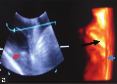


(a) Sonogram of stomach in first case of gastritis thickening of gastric wall (blue arrow). Gastric folds are hyperechoic (red arrow in two-dimensional [2D] sonogram and black
arrow in three-dimensional [3D] sonogram),
(b) sonogram of stomach in second case of gastritis thickening of gastric wall (red arrow in 2D sonogram and black arrow
in 3D sonogram),
(c) sonogram of stomach in third case of gastritis thickening of gastric wall (black arrow),
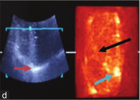
(d) sonogram of stomach fourth case of gastritis thickening of gastric wall (red arrow in 2D sonogram),
(e) sonogram of stomach in fifth
case of gastritis thickening of gastric wall (white arrow in 2D sonogram). Gastric fold are hyperechoic (red arrow in 2D
sonogram and green arrow in 3D sonogram), (f) sonogram of stomach in sixth case of gastritis thickening of gastric wall
(green arrow).
Madan Pal, Prem Singh, Rishi Tayal, Dinesh Dehmiwal, S. M. Behl, Sarvan Kumar and R. K. Chandolia: "A comparative study of two-dimensional and three-dimensional
ultrasonography in evaluation of gastric affections in dogs"; Veterinary World, EISSN: 2231-0916


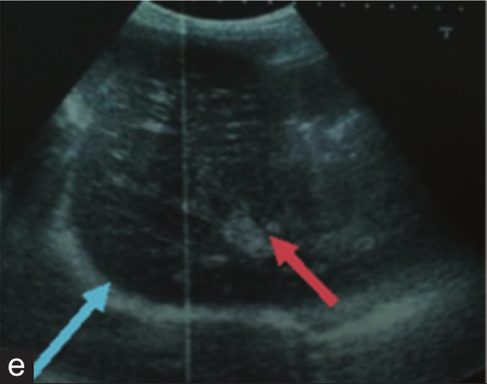

(a) Two-dimensional (2D) sonogram in case of gastric ulcer, thickening of gastric wall (black arrow) and disruption
of mucosal layer is hyperechoic (red arrow), (b) gastric lesion on mucosa of stomach as gastric ulcer,
(c) 2D sonogram in first case of gastric dilatation, there is anechoic fluid (blue arrow) in lumen of stomach due to whick boundaries are not clear,
(d) 2D sonogram in second case of gastric dilatation the solid food in lumen of stomach is hyperechoic (red arrow)
and there is dilatation of stomach due to which boundaries of stomach is not clear, (e) 2D sonogram in third case of gastric
dilatation, there is shadow of full sized stomach due to dilatation with the solid food in lumen of stomach is hyperechoic
(red arrow) and thickening of gastric wall as hyperechoic shadow (blue arrow),
(f) 2D sonogram of gastric foreign bodies,
there is hyperechoic shadow of bunch of straw (red arrow) and leather piece (green arrow) in lumen of stomach.
Madan Pal, Prem Singh, Rishi Tayal, Dinesh Dehmiwal, S. M. Behl, Sarvan Kumar and R. K. Chandolia: "A comparative study of two-dimensional and three-dimensional
ultrasonography in evaluation of gastric affections in dogs"; Veterinary World, EISSN: 2231-0916


(a) Two-dimensional sonogram of pyloric
stenosis, there is hyperechoic thickening of muscle of pyloric antrum (red arrow),
(b) three-dimensional sonogram of
pylori stenosis, there is hyperechoic thickening of muscle of pylori antrum (black arrow).
Madan Pal, Prem Singh, Rishi Tayal, Dinesh Dehmiwal, S. M. Behl, Sarvan Kumar and R. K. Chandolia: "A comparative study of two-dimensional and three-dimensional
ultrasonography in evaluation of gastric affections in dogs"; Veterinary World, EISSN: 2231-0916
