

Article added / Artikel hinzugefügt 01.10.2021
Generally Articles and Discussions about Osteosarcoma in Dogs
→ Evaluations of phylogenetic proximity in a group of 67 dogs with
osteosarcoma: a pilot study
Article added / Artikel hinzugefügt 01.10.2021
Generally Articles and Discussions about Osteosarcoma in Dogs
→ Canine Periosteal Osteosarcoma
Images added / Abbildungen hinzugefügt 02.05.2019
Generally Sonography Atlas of Dogs →
Cardiovascular system → Pulmonary vessels
New subcategory added / Neue Unterkategorie hinzugefügt 02.05.2019
Generally Sonography Atlas of Dogs →
Cardiovascular system → Pulmonary vessels
Images added / Abbildungen hinzugefügt 01.05.2019
Generally Sonography Atlas of Dogs →
Cardiovascular system → Heart valvular diseases


Generally Sonography Atlas of Dogs
(Allgemeiner Sonographie-Atlas von Hunden)
Cardiovascular system - Liver vessels
(Kardiovaskuläres System - Lebergefäße)

Spectral trace from a German Shepherd dog with portal hypertension due to a pancreatic carcinoma

Different velocity profile retrieved from the same patient using the right angle correction (left) and the wrong one (right)
Pablo Gomez Ochoa, Delia Lacasta, Ivan Sosa, Manuel Gascon, Juan Jose Ramos and Luis Miguel Ferrer (2011). Foundamentals and Applications of Abdominal Doppler, Ultrasound Imaging - Medical Applications, Prof. Oleg Minin (Ed.), ISBN: 978-953-307-279-1, InTech, DOI: 10.5772/20333. Available from: http://www.intechopen.com/books/ultrasound-imaging-medical-applications/foundamentals-and-applications-of-abdominal-doppler


Figure 13: Tortuous vessels corresponding to nephro-splenic shunts in a dog with cirrhosis
Pablo Gomez Ochoa, Delia Lacasta, Ivan Sosa, Manuel Gascon, Juan Jose Ramos and Luis Miguel Ferrer (2011). Foundamentals and Applications of Abdominal Doppler, Ultrasound Imaging - Medical Applications, Prof. Oleg Minin (Ed.), ISBN: 978-953-307-279-1, InTech, DOI: 10.5772/20333. Available from: http://www.intechopen.com/books/ultrasound-imaging-medical-applications/foundamentals-and-applications-of-abdominal-doppler


Portal hypertension in a caval syndrome. The image shows the congestive liver, huge hepatic veins related to portal vessels.
Pablo Gomez Ochoa, Delia Lacasta, Ivan Sosa, Manuel Gascon, Juan Jose Ramos and Luis Miguel Ferrer (2011). Foundamentals and Applications of Abdominal Doppler, Ultrasound Imaging - Medical Applications, Prof. Oleg Minin (Ed.), ISBN: 978-953-307-279-1, InTech, DOI: 10.5772/20333. Available from: http://www.intechopen.com/books/ultrasound-imaging-medical-applications/foundamentals-and-applications-of-abdominal-doppler


a: Sonographic study of the liver. There is distention of the intrahepatic portal vein system. The cross-sectional diameter of the portal vein at the porta hepatis measured 0.92 cm.
The diameter of the aorta at the level of the diaphragm measured IS 0.77 cm. The liver has a slightly patchy echoic pattern. b: The walls of the hepatic veins are small, hyperechoic,
and thickened relative tothe intralobar portal veins.
Cohn, L. A., Spaulding, K. A., Cullen, J. M., Bunch, S. E., Metcalf, M. R., Hardie, E. M., MacLachlan, N. J. and Breitschwerdt, E. B. (1991), Intrahepatic Postsinusoidal Venous Obstruction in a Dog. Journal of Veterinary Internal Medicine, 5: 317–321. doi: 10.1111/j.1939-1676.1991.tb03144.x


Ultrasonographic image of liver with engorged portal veins (PV) and hepatic veins (HV).
Kumar KS, Srikala D. "Ascites with right heart failure in a dog:diagnosis and management". www.scopemed.org/?mno=157163 [Access: September 23, 2016]. doi:10.5455/javar.2014.a15


Dorsal sonogram (2D) of liver depicting hypoechoic parenchyma with portal vessel dilatation in one-year-old mixed-breed dog.
Kumar V, Kumar A, Varshney AC, Tyagi SP, Kanwar MS, Sharma SK. Diagnostic Imaging of Canine Hepatobiliary Affections: A Review. Veterinary Medicine International. 2012;2012:672107. doi:10.1155/2012/672107.


Longitudinal sonogram (2D) of liver depicting hypoechoic parenchyma with marked portal vessel dilatation (subjective) in 5-year-old mixed-breed dog with significant spleenomegaly.
Kumar V, Kumar A, Varshney AC, Tyagi SP, Kanwar MS, Sharma SK. Diagnostic Imaging of Canine Hepatobiliary Affections: A Review. Veterinary Medicine International. 2012;2012:672107. doi:10.1155/2012/672107.

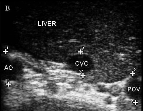
The transverse view of normal canine porta hepatis. Three major vessels in a dorsoventral direction are abdominal aorta (AO), the most dorsal structure, caudal vena cava (CVC) view at margin of the liver and the portal vein (POV) located ventrally (dorsal is to the left, ventral is to the right, and medial is to the bottom).
WICKRAMASEKARA RAJAPAKSHAGE, B. K., ELLEARAEWE GARUHAMILAGE, J. P. K., DE SILVA, D. D. N., & DANGOLLA, A. (2016). "Dimensional ultrasonographic relationship of the right lobe of pancreas with associated anatomic landmarks in clinically normal dogs". http://doi.org/10.1292/jvms.15-0209
