

Article added / Artikel hinzugefügt 01.10.2021
Generally Articles and Discussions about Osteosarcoma in Dogs
→ Evaluations of phylogenetic proximity in a group of 67 dogs with
osteosarcoma: a pilot study
Article added / Artikel hinzugefügt 01.10.2021
Generally Articles and Discussions about Osteosarcoma in Dogs
→ Canine Periosteal Osteosarcoma
Images added / Abbildungen hinzugefügt 02.05.2019
Generally Sonography Atlas of Dogs →
Cardiovascular system → Pulmonary vessels
New subcategory added / Neue Unterkategorie hinzugefügt 02.05.2019
Generally Sonography Atlas of Dogs →
Cardiovascular system → Pulmonary vessels
Images added / Abbildungen hinzugefügt 01.05.2019
Generally Sonography Atlas of Dogs →
Cardiovascular system → Heart valvular diseases


Generally Sonography Atlas of Dogs
(Allgemeiner Sonographie-Atlas von Hunden)
Cardiovascular system - Heart
(Kardiovaskuläres System - Herz)

Simultaneous M-mode and pulse-wave Doppler recording of mitral E and A waves.
Pereira, Guilherme G., Petrus, Liliam C., Santos, André L.F., Yamaki, Fernanda L., & Larsson, Maria Helena M.A.. (2009). "Evaluation of left ventricular diastolic echocardiographic parameters in healthy dogs by pulsed-wave Doppler." Pesquisa Veterinária Brasileira, 29(4), 291-294. Retrieved February 21, 2016, from http://www.scielo.br/scielo.php?script=sci_arttext&pid=S0100-736X2009000400003&lng=en&tlng=en.


Pulse-wave Doppler recording of IVRT.
Pereira, Guilherme G., Petrus, Liliam C., Santos, André L.F., Yamaki, Fernanda L., & Larsson, Maria Helena M.A.. (2009). "Evaluation of left ventricular diastolic echocardiographic parameters in healthy dogs by pulsed-wave Doppler." Pesquisa Veterinária Brasileira, 29(4), 291-294. Retrieved February 21, 2016, from http://www.scielo.br/scielo.php?script=sci_arttext&pid=S0100-736X2009000400003&lng=en&tlng=en.


M-mode echocardiogram of healthy dog - note the lumen of right ventricle (RV), left ventricle (LV).
Kumar KS, Srikala D. Ascites with right heart failure in a dog: diagnosis and management. www.scopemed.org/?mno=157163 [Access: September 25, 2016]. doi:10.5455/javar.2014.a15


Left parasternal short-axis scanning technique. The transducer is positioned near the left cranial border of the heart. Cranial-caudal yields the series of short axis (SAx) images. Variable angulation in the dorsal-ventral direction may be necessary as suggested by the figure. 1: Left parasternal short-axis (LPS SAx) view at the right ventricle (RV) inflow tract. The right coronary (RC), left coronary (LC), and non-coronary (NC) cusps of the aortic (Ao) valve may be visible. 2: LPS SAx view at the RV inflow-outflow tracts. Both the inflow and outflow tracts of the right heart may be visible with subtle angulation and twist adjustments. The RC, LC, and NC cusps of the Ao valve may be visible. 3: LPS SAx view at the pulmonary trunk (PT). Division of the PT into the right and left main pulmonary arteries is seen in the far field.

With special thanks to the authors of the book "Small Animal Ultrasonography" , Marc-André d’Anjou and
Dominique Penninck

Pulsed wave Doppler recording of normal transmitral flow from the left apical 4-chamber view. Early (E) and late (A) diastolic filling waves are noted. Deceleration time of the E wave (DTe)
is
measured as the time from peak E to return to baseline.
O'Sullivan, M. L., O'Grady, M. R. and Minors, S. L. (2007), Assessment of Diastolic Function by Doppler Echocardiography in Normal Doberman Pinschers and Doberman Pinschers with Dilated Cardiomyopathy. Journal of Veterinary Internal Medicine, 21: 81–91. doi: 10.1111/j.1939-1676.2007.tb02932.x


Pulsed wave Doppler recording of normal pulmonary venous flow from the left caudal lobar pulmonary vein from the left
apical 4-chamber view. Systolic (S), diastolic (D), and atrial reversal (AR) waves are
noted.
O'Sullivan, M. L., O'Grady, M. R. and Minors, S. L. (2007), Assessment of Diastolic Function by Doppler Echocardiography in Normal Doberman Pinschers and Doberman Pinschers with Dilated Cardiomyopathy. Journal of Veterinary Internal Medicine, 21: 81–91. doi: 10.1111/j.1939-1676.2007.tb02932.x


Pulsed-wave Doppler recording of normal lateral mitral annular motion. Sample volume position and alignment are seen in the inset 2-Dimensional image of the
left atrium and ventricle from the left apical window. Systolic (Sm), early diastolic (Em), and latediastolic (Am) waves are noted.
O'Sullivan, M. L., O'Grady, M. R. and Minors, S. L. (2007), Assessment of Diastolic Function by Doppler Echocardiography in Normal Doberman Pinschers and Doberman Pinschers with Dilated Cardiomyopathy. Journal of Veterinary Internal Medicine, 21: 81–91. doi: 10.1111/j.1939-1676.2007.tb02932.x

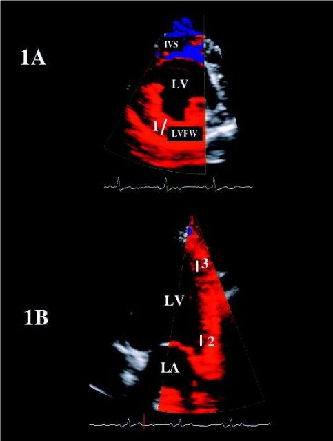
Two-dimensional color TDI images obtained from the right parasternal short-axis view (1A) and
the left apical 4-chamber view (1B) for radial and longitudinal strain and strain rate imaging of the
left ventricular free wall respectively. White bars (1 to 3) represent sample segments located within the left ventricular free wall thickness (1) for the radial motion, and at the base (2)
and the apex (3) of the same wall for the longitudinal motion. IVS: interventricular septum. LA: left atrium. LV: left ventricle. LVFW: left ventricular free wall
Valerie Chetboul, Carolina Carlos Sampedrano, Vassiliki Gouni, Audrey P. Nicolle, Jean-Louis Pouchelon, and Renaud Tissier
"Ultrasonographic Assessment of Regional Radial and Longitudinal
Systolic Function in Healthy Awake Dogs"
DOI: 10.1111/j.1939-1676.2006.tb01802.x


Herzultraschall eines Hundes
Mit Dank an die Tierärztlichen Spezialisten in Hamburg für die freundliche Unterstützung: http://www.tsh.de


Hundeherz, 2D/M-Mode. Die Bewegung des Herzmuskels wird entlang der senkrechten Linie im (oberen) 2D-Bild im unteren M-Bereich aufgetragen
https://de.wikipedia.org/wiki/Sonografie
https://upload.wikimedia.org/wikipedia/commons/9/9c/B_m-mode1.jpg


Gesundes Hundeherz, Dachshund-Mix 4 Jahre / Healthy heart of a 4 yrs. old dachshound-mix
