

Article added / Artikel hinzugefügt 01.10.2021
Generally Articles and Discussions about Osteosarcoma in Dogs
→ Evaluations of phylogenetic proximity in a group of 67 dogs with
osteosarcoma: a pilot study
Article added / Artikel hinzugefügt 01.10.2021
Generally Articles and Discussions about Osteosarcoma in Dogs
→ Canine Periosteal Osteosarcoma
Images added / Abbildungen hinzugefügt 02.05.2019
Generally Sonography Atlas of Dogs →
Cardiovascular system → Pulmonary vessels
New subcategory added / Neue Unterkategorie hinzugefügt 02.05.2019
Generally Sonography Atlas of Dogs →
Cardiovascular system → Pulmonary vessels
Images added / Abbildungen hinzugefügt 01.05.2019
Generally Sonography Atlas of Dogs →
Cardiovascular system → Heart valvular diseases


NORMAL OCULAR ULTRASONOGRAPHIC FINDING IN DOG

Mehdi Tavana and Seyedeh Zeinab Peighambarzadeh, "NORMAL OCULAR ULTRASONOGRAPHIC FINDING IN DOG", www.cibtech.org/sp.ed/jls/2014/03/jls.htm

ABSTRACT
Ultrasonography is a relatively easy , safe and non -invasive examination method which can be used in
diagnosis of ocular disorders as complementary to routine ophthalmic examinations especially when
severe swelling of the eyelid, keratitis, cataract and intraocular hemorrhage prevent direct ophthalmic
examinations. Transcorneal ultrasonographic scanning of left and right eyes of 10 dogs (5 male and 5
female) was performed using a 7.5 -10 MHz transducer. Qualitative ultrasonographic findings of the eyes
were described and measurements of the ocular structures were obtained. Mean ±standard deviation of the
anterior -posterior length of the eye axis, thickness of the lens and depth of the anterior chamber were as
19.41±0.78, 5.71±0.45 and 8.63±0.35 mm ,respectively. The result of this study can be used as normal
value measurements in ultrasonographic evaluation of mixed breed dog 's eye .
Keywords: Ocular , Ultrasonographic Finding , Dog
INTRODUCTION
Ultrasonography is an important method for the clinical assessment of various ocular and orbital diseases.
With understanding of the indications for ultrasonography and proper examination technique, one can
gather a vast amount of information not possible with clinical examination alone. B-scan ultrasound is
most useful when direct visualization of intraocular structures is difficult or impossible. Ultrasonography
has been used since 1956 for diagnostic ocular diseases in humans. Veterinary ocular ultrasonography
was first described in 1968 (Coile and O’Keefe, 1998). Situations that prevent normal examination
include lid problems (eg, severe edema, partial or total tarsorrhaphy), keratoprosthesis, corneal opacities
(eg, scars, severe edema), hyphema, hypopyon, miosis, pupillary membranes, dense cataracts, or vitreous
opacities (eg, hemorrhage, inflammatory debris) (Qureshi et al., 2010).
Corneal edema is a common clinical sign of corneal ulceration, keratitis, anterior uveitis, and many
systemic diseases, and precludes the direct visualization of intraocular structures by ophthalmoscopy
(Whittaker et al., 1999). Under such conditions; alternative diagnostic methods for intraocular diseases
must be explored (Boroffka et al., 1998; Bentley et al., 2003; Scotty et al., 2004).
Ultrasonography is a useful tool to evaluate the contents of the globe and orbit as is done routinely in
companion animal medicine (Ribeiro et al., 2009). Ultrasonography is used for: (a) evaluation of
intraocular details obscured from visualization by the ocular media opacities. (b) Evaluation of
retinochoroidal lesions especially tumors even with clear media (c) differentiation of solid from cystic and
homogenous from heterogeneous masses. (d) Examination of retrobulbar soft tissue masses and normally
present orbital structures (to differentiate proptosis from exophthalmos). (e) Identification, localization
and measurement of non radio-opaque/radio-opaque foreign bodies. (f) Biometry and pachmetry. (g)
Follow up evaluations.
Transcorneal ultrasonography enables the evaluation of intraocular structures in eyes with opaque,
diseased corneas in order to evaluate the prognosis for vision following resolution of the corneal disease.
The evaluation of ocular emergencies can be limited by lack of sophisticated tools and training. Direct
visualization of intraocular structures is difficult or impossible when the eye lids are swollen shut after
injury. Lens opacification and hyphema can also block the posterior view of the chamber.
Indications for ocular ultrasound include any clinical entity which impedes visualization of the globe and
retrobulbar region. Severe corneal edema, corneal lacerations or ulcerations, cataracts or ocular masses
may preclude visualization of deeper structures with traditional ophthalmoscopic methods. Another
common indication for ocular ultrasound is disparity in globe size or an exophthalmic globe.
Knowledge of the ultrasonographic appearance and normal dimensions of the eye would be serve as a
basis for ultrasonographic examinations when ocular disease may have caused alterations in dimensions
and appearance. The aim of this study was to measure and evaluate the structures of the eye (the thickness
of the cornea, lens, and vitreous, and sagital eyeball axis) in adult mixed- breed dogs using ultrasound
equipment.
MATERIALS AND METHODS
Ten clinically healthy adult mixed- breed dogs, with no evidence of ocular disorder, five male and five
female, weighing 8 – 10 kilograms, were prepared. All dogs had a pre-study ophthalmic examination that
demonstrated dog’s eyes were normal. Dogs were sedated intramuscularly with mixture of Xylazin and
Diazepam.
The left and right eyes of dogs were scanned ultrasonographically in using a Piomedical ultrasound
machine with a linear array transducer of 7.5-10 MHz.
To avoid trapping air between the transducer and the patient, the palpebral hair was thoroughly wetted
before the acoustic gel was applied. After using enough gel on the upper eyelids, transcorneal scanning
was started while the globe was imaged in both horizontal and vertical planes through the visual axis on
the upper eyelid and perpendicular to the upper palpebral fissure respectively for a complete examination.
This produces a cross-sectional image of the eye with the medial canthus to the right and lateral canthus
to the left of the horizontal images. Attempts were made not to cause pressure on the cornea during
placement of the transducer on the eyelid. The gain was set so that there was an anechoic region between
the anterior and posterior lens capsule.
We measured five points in the adult dog’s cornea: central, peripheral superior, peripheral inferior,
peripheral nasal and peripheral temporal. The peripheral points were 1 mm from limbus (the junction of
cornea and sclera). Ten measurements were taken at each point to determine the mean thickness. A
Student t-test was used for statistical analysis.
RESULTS AND DISCUSSION
Results
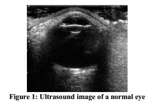
In B-mode ultrasonography, four major echoes include cornea, anterior lens capsule, posterior lens
capsule and retina-choroid sclera complex were easily seen (Figure 1). The cornea was represented as a
curved hyper parallel echoic interface immediately under the eyelid. In some of the sonograms, the cornea
could be seen as three thin layers, in which the anterior and posterior layers were quite hyper echoic and
the middle layer appeared anechoic. Anterior chamber of the eye, lens cortex and nucleus and the vitreous
were anechoic. Other echoic structures including Ciliary body, iris and optic disc could also be
distinguished.
The lens thickness ranged from 4.9 to 7.1mm in adult dogs. The vitreous thickness ranged respectively
from 18.8 to 20.9 mm. The parameters of the left and right eye differed insignificantly. The thinnest
central cornea was 0.50±0.12 mm in the left eye and 0.58±0.13 mm in the right eye. Corneal thickness
ranged from 0.36 to 0.73 mm. The central part of the cornea was the thinnest and peripheral part was
slightly thicker in adult dog. The peripheral temporal point of the cornea was the thickest. The results of
the measurements on dog’s eye were recorded in Table 1.

Discussion
Ocular ultrasound is an addition to, not a replacement for, routine ophthalmic examination including
assessment of menace, blink and papillary light response, fluorescein staining, nasolacrimal evaluation,
determination of intraocular pressure and examination of anterior and posterior segments using a bright
focal light source and direct and indirect ophthalmoscopy or bio microscopy, respectively (Reef, 1998;
Gonzlez et al., 2001). Several studies have been done on the eye ultrasonography in different kind of
animals Veterinary (Coile and O’Keefe, 1998; Hughes, 1972; Hughes 1979). The most common clinical
indications for ultrasound are to evaluate for the presence of a retinal detachment in eyes with a cataract,
intraocular lesions including lens displacement, intravitreal hemorrhage and intraocular foreign bodies. In
addition, orbital evaluation can be performed in instances of exophthalmoses or orbital trauma (Hillyer,
1993). In the study reported here anterior-posterior length of the eye axis, lens diameter, depth of the
anterior chamber and depth of vitreous were measured in normal eyes of dogs to establish mean and
standard deviation values. No difference was detected in any ocular component measurement between the
right and left eyes of the adult mixed-breed dogs. It was in agreement with other investigations similar
studies on enucleated eye of dog have been done.
REFERENCES
Bentley E, Miller PE and Diehl KA (2003). Use of high-resolution ultrasound as a diagnostic tool in
veterinary ophthalmology. Journal of American Veterinary Medicine Association 223 1617-1622.
Boroffka S, Verbruggen AM, Boevé MH and Stades FC (1998). Ultrasonographic diagnosis of
persistent hyperplastic tunica vasculosa lentis /persistent hyperplastic primary vitreous in two dogs.
Veterinary Radiology and Ultrasound 39 440-444.
Coile DC and O’Keefe LP (1998). Schematic eyes for domestic animals. Ophthalmic and Physiological
Optics 8 215-220.
Hillyer MH (1993). Ocular ultrasonography in the horse In: The Veterinary Annual, 33rd edition, edited
by Raw ME and Parkinson (Oxford, Blackwell Scientific Publications) 131-137.
Hughes A (1972). A schematic eye for the rabbit. Vision Research 12 123-138.
Hughes A (1979). A schematic eye for the rat. Vision Research 19 569-588.
Gonzalez EM, Rodriguez A and Garcia I (2001). Review of ocular ultrasonography. Veterinary
Radiology and Ultrasound 42 485-495.
Qureshi MA and Laghari K (2010). Role of B-scan ultrasonography in pre-operative cataract patients.
International Journal of Health Scienceses (Qassim) 4(1) 31-7.
Reef VB (1998). Equine diagnostic ultrasound Philadelphia, Pennsylvania (WB Saunders Company) 481-
536.
Ribeiro AP, Silva ML, Rosa JP, Souza SF, Teixeira IA and Laus JL (2009). Ultra-sonographic and
echobiometric findings in the eyes of Saanen goats of different ages. Veterinary Ophthalmology 12 313-
317.
Scotty NC, Cutler TJ, Brooks DE and Ferrell E (2004). Diagnostic ultrasonography of equine lens and
posterior segment abnormalities. Veterinary Ophthalmology 7 127-139.
Whittaker CJG, Gelatt KN and Wilkie DA (1999). Food animal ophthalmology In: Veterinary
Ophthalmology, 3rd edition, edited by Gelatt KN (Lippincott Williams & Wilkins) Philadelphia, PA
1117- 1176.
Share this article / Teilen Sie diesen Artikel