

Article added / Artikel hinzugefügt 01.10.2021
Generally Articles and Discussions about Osteosarcoma in Dogs
→ Evaluations of phylogenetic proximity in a group of 67 dogs with
osteosarcoma: a pilot study
Article added / Artikel hinzugefügt 01.10.2021
Generally Articles and Discussions about Osteosarcoma in Dogs
→ Canine Periosteal Osteosarcoma
Images added / Abbildungen hinzugefügt 02.05.2019
Generally Sonography Atlas of Dogs →
Cardiovascular system → Pulmonary vessels
New subcategory added / Neue Unterkategorie hinzugefügt 02.05.2019
Generally Sonography Atlas of Dogs →
Cardiovascular system → Pulmonary vessels
Images added / Abbildungen hinzugefügt 01.05.2019
Generally Sonography Atlas of Dogs →
Cardiovascular system → Heart valvular diseases


Spontaneous Osteosarcoma in Dogs: Diagnosis through Cytopathological and Histopathological Assays

Gomes MC, Rocha NS (2016) "Spontaneous Osteosarcoma in Dogs:
Diagnosis through Cytopathological and Histopathological Assays".
J Cytol Histol 7: 404. doi:10.4172/2157-7099.1000404

Abstract
Background: Appendicular osteosarcoma is the main primary malignant and non-hematopoietic bone tumor in dogs. It appears spontaneously in the appendicular skeleton with a
relevant incidence both in dogs and in human children. Unfortunately, it is an aggressive neoplasm with high rates of metastasis regardless of the species and that afflicts, among other organs,
the lungs. The high rate of lethality is due to the terrible survival prognosis for patients with lung metastasis and due to chemotherapy. Traditionally, amputation is the therapeutic indication,
followed by chemotherapy. For human patients, the chemotherapeutic treatment, based on a prior anatomopathological diagnosis referencing even the degree of aggressiveness, results in a better
rate of survival as it reduces metastases. However, this approach is not often explored in veterinary medicine, which features amputation as the traditional approach, followed by
chemotherapy.
Objective: Verify the morphological expression of the parameters used in the cytopathological diagnosis through fine needle aspiration cytology of spontaneous osteosarcomas in
dogs from varying breeds, including mixedbreeds. The study also aims the analysis of possible concordances between cytopathological and histopathological parameters in osteosarcomas.
Methods: This study verified, through optical microscopy, the morphological expression of parameters used in the cytopathological diagnosis of osteosarcomas in twenty dogs of
different breeds, including mixed-breeds, and then conducted an analysis of possible concordances between these parameters. The cytological assay was conducted through fine needle aspiration
cytology, using Giemsa and Papanicolaou stain on the microscope slides. The histopathological assay was conducted through the use of biopsies of surgical specimens, which were processed according
to the standard procedures and stained with hematoxylin-eosin. The tumors were classified based on the guidelines proposed by the World Health Organization.
Results: There were malignancy criteria significantly repeated both at cytopathological and at histopathological assays.
Conclusion: These findings show us that the cytopathological assay through aspiration may be used as a trustworthy diagnostic method for osteosarcomas in dogs.
Keywords
Osteosarcoma; Dogs; Cytopathology; Histopathology
Introduction
Osteosarcomas (OSAs) or osteogenic sarcomas are, among bone neoplasms, the primary tumors with the highest incidence in dogs, as well as in human children, despite being about ten times more frequent in the canine species. This biological similarity has been turning dogs into a clinic model for the study of this type of cancer in humans [1,2], with several advantages over the study model in mice [3].
Most OSAs in dogs are malignant, as they may usually cause death through local infiltration, with about 80% of the dogs afflicted by OSA dying due to pulmonary metastases [4]. Over 15% of the patients with the clinical symptomatology are detected with pulmonary metastasis and it is estimated that over 80% present micrometastases [5].
Characteristically, OSA is found in the metaphyses of long bones and in the appendicular skeleton, with about 25% of the cases in dogs affecting the axial skeleton [6]. This neoplasms appear primarily in dogs with long limbs, such as the Irish Wolfhound, the Scottish Wolfhound and the Great Dane breeds [2,7,8], as well as in other large and giant breeds such as St. Bernard, Irish Setter, Dobermann, Rottweiler, German Shepherd and Labrador Retriever [2,6,8,9], including mixedbreeds [3,10,11].
OSAs afflict dogs from middle to old age, with an average age of seven to eight years old. Moreover, males tend to be afflicted more than females, but this statement is not a consensus among researchers, and some studies have not noticed any gender predisposition [8,12].
The biologic behavior of OSAs is an aggressive local infiltration of the adjacent tissues and a fast hematogenic spread, usually to the lungs. Appendicular OSAs usually appear in the distal radius metaphysis, in the distal femur metaphysis and in the proximal humerus metaphysis, although other metaphyses may also be affected [13].
At the imaging exams, we can observe a mixed lytic-proliferative pattern at the metaphysis of the afflicted bone and an adjacent periosteal bone formation, which leads to the development of Codman’s triangle, formed by the cortex of the afflicted area and the periosteal proliferation. Just like in humans, pulmonary metastases are the main causes of terminal morbidity, suggesting that over 90% of the canine patients may present microscopic metastases undetectable on imaging techniques during the routine [14].
Aside from the patient’s clinical history, a detailed physical examination and radiographic examinations, the diagnosis is also based on a cytological assay, with the confirmation often being made through biopsy and histopathological assay [1,15]. The case study representativeness of this tumor is low in the Brazilian territory, due mainly to owners’ choice for euthanasia given the high cost of the treatment. Therefore, records regarding the disease and other information regarding its clinic and pathologic manifestations are lost, both ante and post-mortem [14,16].
According to Ribeiro et al. [17], the early diagnosis of jaw OSA in humans favorably influences the treatment and prognosis of the disease, as, with a fast diagnosis and a precise assessment of the tumoral involvement, it is possible to conduct a conservative treatment with curative goals and minimal sequelae.
The treatment of OSA consists of amputation or limb-sparing surgery followed with adjuvant chemotherapy with doxorubicin, platinum-based drugs, or a combination of both, as well as cisplatin and carboplatin. The average survival of these animals with amputation and chemotherapy without metastasis ranges from 165 to 470 days [4,18- 21]. With advances in the treatment and multi-agent chemotherapy, the prognosis has improved during the last few decades, with an increased survival rate, but the prognosis remains bad for patients with pulmonary metastasis or patients with refractory tumors [5].
Canine OSAs share many traits with human OSAs, including wounds of identical appearance, with dogs possibly being used as a comparative model [13,14]. Given the frequency in dogs, the canine model for spontaneous OSA has been offering unique opportunities towards comprehending the genomic origins of this tumor. This allows both studies regarding the role of metastases in the disease and tests with new research drugs that would otherwise take too long to provide results in humans [9].
Regarding morphological microscopic characterization, OSA cells are usually round or elliptical, with defined cytoplasmic borders, a bright blue granular cytoplasm and eccentric nucleus with or without nucleoli. Giant, multinucleated cells are common and often there is an amorphous pinkish material (osteoid) at the slide background or in the osteoblastic cytoplasm [22,23]. If the round cells cannot be identified with confidence as osteoblasts, it is possible to conduct, in non-stained slides, a cytochemical stain for alkaline phosphatase (ALP) as osteoblasts are usually ALP positive [15].
The Fine Needle Aspiration Cytology (FNAC) was created in the 1930s with the purpose of diagnosing malignant tumors in humans. In animals, the technique began to be employed in the 1980s, aiding in the distinction between hyperplasia, inflammations, neoplasms and degenerations [23-26]. OSA FNAC is usually conducted using a bone marrow aspiration needle. In most cases, a blunt percutaneous FNAC may be conducted with only manual containment – if the operator cannot penetrate the cortex, the ultrasound guide usually allows the visualization of a “window” through which the needle is inserted. The FNAC method potentially perfects the cytological sample due to architectural preservation, allowing the creation of paraffin blocks for later processing, similarly to a histological sample, which enables the use of histochemical and immunohistochemical adjuvant techniques [27].
Although this approach is seldom explored in veterinary medicine, the role of cytology as a diagnostic tool continues to expand [1]. The technique has several advantages, such as reliability, a minimally invasive diagnosis, reduced cost in comparison to histopathology and fast results [22,28], which enables the surgical and therapeutic approach. Despite the more specific and definitive characteristics related to the diagnosis through histopathology, several authors consider that cytopathology may be used as a definitive diagnosis [23,29-31], or at least be considerably helpful [32]. However, there are still several restrictions regarding the sensitivity of this method [25], as the irregular staining and the presence of precipitate or other refracting artifacts [26].
Ultimately, both techniques continue to be used in complementary diagnoses, illustrating an option between the low degree of invasion during sample collection for cytopathology and the higher level of information available to assess tissue architecture for histopathology [22].
This study aimed at verifying the morphologic expression of parameters used in the cytopathological diagnosis of spontaneous OSAs in dogs of several breeds, including mixed-breeds. It also aimed at analyzing the possible concordance between cytopathological and histopathological parameters of these bone neoplasms.
Materials and Methods
For this experiment, we used canine OSAs diagnosed at the Veterinary Hospital and Veterinary Pathology Service at FMVZ – UNESP, Botucatu Campus, Brazil. Patients have, in order to reach a diagnosis, undergone clinical, radiologic, surgical and pathological examinations. The presumptive diagnosis was reached by cytological findings and confirmed by histopathological findings, as recommended by the World Health Organization – Histological Classification of Bone and Joint Tumors of Domestic Animals [32].
All owners received explanations regarding the procedures of this study, signing a Free, Prior and Informed Consent term. The project was approved in a favorable decision at the Ethics Council at FMVZ – UNESP, Botucatu Campus, Brazil.
The samples were collected from dogs with defined breeds and mixed-breeds with definitive diagnosis of OSA, totaling twenty animals. These animals firstly underwent FNAC of the lesion, followed by excision of the tumor, cytopathological and histopathological slides processing and their reading. The final diagnosis of OSA was given by distinct pathologists, and the slides were then archived in our Department of Veterinary Pathology. They were once more analyzed in behalf of this study.
In the cytopathological exam, each tumor was firstly divided in four quarters, followed by FNAC of each one. At least three microscope slides were used by quarter. The slides were first fixed through methanol and then stained by Giemsa; in those stained by Papanicolaou, 95% alcohol was used instead. All these proceedings were performed by different operators.
Surgical specimens were obtained from animals that underwent amputation or tumor excision. The tumor fragments were fixated in 10% neutral buffered formalin by at least 72 hours. They were then decalcified in 10% nitric acid and processed by the usual histological techniques of dehydration, diafanization and embedded in paraffin, cut with a maximum thickness of four micrometers, and stained with Hematoxylin-Eosin.
In this study, we first verified the quality of the samples, observing staining pattern and cellularity. Then, we then detailed the cellular characteristics for the diagnosis of OSA using previously established malignancy criteria for neoplasms as recommended by the World Health Organization – Histological Classification of Bone and Joint Tumors of Domestic Animals [33].
Results
Twenty dogs participated in this study, with thirteen pure breeds (65%) and seven mixed breeds (35%). The average age of the animals was 9.5 years old, and the average weight was 34.8 kg (Tables 1 and 2).
Table 1: Information regarding weight, gender and age of the twenty dogs with OSA.

Table 2: Percentage of each breed affected with OSA, based on the thirteen pure breeds found in this study (65% of the twenty animals). There were seven (35%) mixed breed dogs in this study.

In the present study, we have analyzed the morphological expression of several diagnostic parameters for OSA in dogs for comparison between diagnosis techniques through cytopathology and histopathology. The malignancy criteria included cellularity (low, moderate and high) and cellular arrangement (isolated or cohesive), as well as characteristics of the nucleus: presence of a nuclear halo, uniform hyperchromasia, prominent and single nucleolus versus multiple nucleoli, nuclear molding, pseudoinclusion, multinucleation, atypical mitosis, enlarged nuclei (karyomegaly), chromatin aspect (finely or coarsely aggregated), presence of macronucleoli and nuclear morphology (rounded, oval and fibrillate). The criteria also consider cytoplasmic characteristics such as broad or scarce cytoplasm, presence of vacuoles and pseudopodia, cannibalism, basophilia, eosinophilia and cytoplasmic morphology (fibrillate or fusiform); anisocytosis and anisocariosis (discrete, moderate or marked); environmental characteristics such as inflammation, immature or mature bone matrix; distribution of mesenchymal cells (chondrocytes, osteoblasts and osteoclasts) and presence of platelets. Some of the aforementioned criteria are represented in (Figures 1 and 2).
Figure 1: Dog osteosarcoma: atypical osteoclast (long white arrow), evident nucleoli (short white arrow), cytoplasm vacuolization (short black arrow), large nucleus-cytoplasm ratio (long black arrow), pseudopodium (gray arrow). Giemsa / 20 μm.
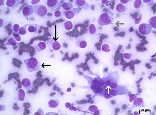
Figure 2: Dog osteosarcoma: disorganized bone trabeculae (short arrow) and atypical osteoclast (long arrow). Hematoxylin-Eosin / 50 μm.

We first conducted a quality test on the microscope slides, with none being considered inadequate for analysis. The staining techniques used in the cytology slides were Giemsa and Papanicolaou.
Criteria such as nuclear halo and pseudo inclusion have not been found in a relevant number, as occurred with criteria such as cannibalism and presence of fibroblasts in the microenvironment.
Due to the low number of cases with an OSA diagnosis in the routine of the veterinary hospital at FMVZ – Botucatu, the number of histopathology slides available for the study did not reach twenty, limiting the comparison study between histopathology and cytopathology to seven cases.
The same steps taken in the examination of the cytopathology slides of OSAs have been employed for the histopathology slides, first with a quality examination followed by an analysis of the malignancy criteria. All samples were previously stained with Hematoxylin-Eosin (Table 3).
Table 3: Anatomical parts afflicted by OSA of twenty dogs. It shows the percentage relating each affected part to its corresponding category (appendicular or axial skeleton).

Due to the nature of the technique employed in the creation of the histopathology slides, it was not possible to clearly establish the criteria related to the cytoplasm, which is much more marked in cytopathological assays with Giemsa staining. Therefore, the analysis of cytoplasmic vacuolization and of the nucleus-cytoplasm ratio were not included. What could be noticed was fibrillate cytoplasmic shape in 57% (4) of the cases and eosinophilia in 85.7% (6).
We have noted a discrepancy between the diagnosis in the system and the findings of this study. Two cases previously diagnosed as osteoblastic OSAs were, in fact, chondrosarcomas, due to the large number of pleomorphic chondrocytes and the marked proliferation of the cartilaginous matrix, characteristics of this kind of neoplasm. Another diagnostic discrepancy was a wound that was revealed a bone remodeling process, in which cells did not present the expected malignancy criteria such as disorganized proliferation of the bone matrix and evident nucleolus.
This study observed that some malignancy criteria for canine OSAs are present both in cytopathology and in histopathology. Upon analyzing (Figures 3 and 4), we may note that the prominent nucleolus criteria is frequent in both techniques (95% in cytology and 85.7% in histology). The same happens with multinucleation (95% and 71.4%, respectively); fibrillate nucleus (50% and 42.9%), high number of osteoblasts (90% and 100%), high number of osteoclasts (85% and 85.7%) and mature bone matrix (85% and 85.7%).
Figure 3: Percentage of the malignancy criteria analyzed in cytopathology slides of osteosarcoma.

Figure 4: Percentage of the malignancy criteria analyzed in histopathology slides of osteosarcoma.

Discussion
As previously mentioned, only seven histopathologic samples were analyzed instead of the twenty anticipated originally due to their scarcity. One hypothesis explaining this fact is the prominent position of the Veterinary Hospital at FMVZ-UNESP, Botucatu Campus, which is located in near the geographic center of the State of São Paulo, one of the most economically relevant states in Brazil. The hospital receives patients from several nearby regions and is considered a reference hospital for veterinary medicine in the country.
It is possible that owners of animals afflicted by this disease will choose not to conduct the treatment due to the distance between their hometowns and the hospital, transportation difficulties and traveling time, which makes the treatment unfeasible to some owners. Of all cases analyzed, seven (35%) animals belonged to residents of Botucatu. The cities of Rio Claro, Cerquilho and Tatuí presented two cases each (10% each), with the remaining seven belonging to residents of the cities of Atibaia, Brotas, Buri, Jaú, São Manuel, São Paulo and São Pedro (5% each).
We may also hypothesize that there is a chance that the studied OSA cases did not receive a surgical indication: there are situations in which the animal is not in good enough health to undergo a surgical procedure. It is also possible that the owners chose euthanasia instead of the treatment due to the low quality of life caused by metastases in vital organs or situations in which the owner does not authorize the surgery. According to Bersano [16], the non-representativeness of OSA in dogs is justified by the owners’ decision for euthanasia instead of proceeding with the treatment, mainly due to the high costs involved.
According to the literature, large breeds like Rottweilers, Irish Setters and Labrador Retrievers show great predisposition for OSA [2,6,8]. Moreover, OSA has more often afflicted bones in the limbs or long bones, representing 85% of the cases, as well as the metaphyses [6]. In addition, 55% of the affected animals were large-sized and 10% were giant-sized.
Females have been more afflicted than males, with rates of 55% and 45% respectively. Despite the literature stating that males are more afflicted, there is still no clear consensus among researchers in this respect [8,12]. The average age of the dogs in this study was 9.5 years old. According to Vanel [8] and Morello [12], the average age of animals afflicted by OSAs was between seven and eight years old. The discrepancy between literature and this study may be explained by the increase in life expectancy of pets due to several factors. Among them, we can mention the higher availability of vaccines, the increasing awareness of owners regarding the geriatric issues of their pets and an improvement in the nutritional quality of pet foods [21]. Moreover, an early diagnosis of neoplastic diseases through cytopathological and histopathological assays enables increasingly efficient treatments, as well as higher life expectancy of patients [17].
According to Vanel et al. [8], chondrosarcomas are the main differential diagnosis for OSAs in dogs, being the second most common primary bone tumor in dogs, representing up to 10% of the cases. This study has found two cases of chondrosarcoma among six histopathological samples (33%). This difference can be explained due to the limited number of samples in the study, which is not representative of the described reality. Therefore, further studies regarding the incidence of chondrosarcomas in the State of São Paulo, Brazil, would be recommended, perhaps encompassing larger areas in order to reach an adequate representativeness of the population.
The results have shown that the cytopathological assay may indeed be used as a definite diagnosis for spontaneous OSA in dogs, as corroborated by several authors [23,29-31], since the malignancy criteria expressed through this technique coincide with those expressed in histopathological assays for this type of neoplasms, such as the presence of osteoids, giant multinucleated cells and single or multiple evident nucleolus [32,33]. However, in order to emphasize the morphological criteria, further research with large enough sample sizes to achieve representativeness is needed.
Conclusion
According to the results obtained in this study, we may conclude that cytopathological assays are viable as a diagnostic method for canine spontaneous OSA, being an equally adequate method for grading the malignancy of the neoplasm. Such factors allow the surgeon to conduct a more adequate and fast therapeutic protocol, which may significantly improve the prognosis of the animal in the face of a naturally aggressive neoplasm with low survival rates.
Acknowledgements
I thank the support of ISB - UNESP for the realization of this project, FAPESP and CNPq for the financial resources.
References
- Mueller F, Fuchs B, Kazer-Hotz BC (2007) Comparative biology of human and canine osteosarcoma. Anticancer Res 27: 155-164.
- Fenger JM, London CA, Kisseberth WC (2014) Canine Osteosarcoma: a naturally occurring disease to inform pediatric oncology. ILAR J 55: 69-85.
- Shahi MH, York D, Gandour-Edwards R, Withers SS, Holt R, et al. (2015) BMI1 is expressed in canine osteosarcoma and contributes to cell growth and chemotherapy resistance. PloS One 10: e 0131006.
- Szewczyk M, Lechowski K, Zabielska K (2014) What do we know about canine osteosarcoma treatment? Review. Vet Res Commun 39: 61-67.
- Jaffe N (2009) Adjuvant chemotherapy in osteosarcoma: An odyssey of rejection and vindication. Cancer Treat Res 152: 219-237.
- Selvarajah GT, Kirpensteijn J (2010) Prognostic and predictive biomarkers of canine osteosarcoma. Vet J 185: 28-35.
- Davis BW, Ostrander EA (2014) Domestic dogs and cancer research: a breed-based genomics approach. ILAR J 55: 59-68.
- Vanel M, Blond L, Vanel V (2012) Imaging of primary bone tumors in veterinary medicine: Which differences? Eur J Radiol 82: 2129-2139.
- Schiffman JD, Breen M (2015) Comparative oncology: what dogs and other species can teach us about humans with cancer. Philos Trans R Soc B BiolSci 370.
- Trost ME, Kommers GD, Brown CC, Barros CSL, Irigoyen LF, et al. (2012) Primary bone neoplasms in dogs: 90 cases. Pesq Vet Bras 32: 1329-1335.
- Arthur EG, Arthur GL, Keeler MR, Bryan JN (2016) Risk of osteosarcoma in dogs after open fracture fixation. Vet Surg 45: 30–35.
- Morello E, Martano M, Buracco P (2011) Biology, diagnosis and treatment of canine appendicular osteosarcoma: Similarities and differences with human osteosarcoma. Vet J 189: 268-277.
- Osborne TS, Khanna C (2012) A review of the association between osteosarcoma metastasis and protein translation. J Com Pathol 146: 132-142.
- Dernell WS, Ehrhart NP, Straw RC, Vail DM (2007) Tumors of the skeletal system: Withrow and MacEwen’s small animal clinical oncology (4th edn.), Saunders Elsevier, St. Louis, Missouri. pp: 540-567.
- Ryseff JK, Bohn AA (2012) Detection of alkaline phosphatase in canine cells previously stained with Wright-Giemsa and its utility indifferentiating osteosarcoma from other mesenchymal tumors. Vet ClinPathol 41: 391-395.
- Bersano PRO (2011) In vitroexpression of cyclooxygenase-2 (Cox2) for osteosarcoma exposed to a Cox2 selective inhibitor. Doctorate Thesis - UniversidadeEstadualPaulista "Júlio de MesquitaFilho" School of Veterinary Medicine and Animal Science. Botucatu, São Paulo, Brazil.
- Ribeiro ALR, Nobre RM, AlvesJr SM, Souza PARS, Silva Jr NG, et al. (2010) The importance of early diagnosis and an accurate tumoral evaluation in the treatment of mandibular osteossarcoma. RevistaOdontoCiência 25: 319-324.
- Bacon NJ, Ehrhart NP, Dernell WS, Lafferty M, Withrow SJ (2008) Use of alternating administration of carboplatin and doxorubicin in dogs with microscopic metastases after amputation for appendicular osteosarcoma: 50 cases (1999-2006). J Am Vet Med Assoc 232: 1504-1510.
- Spodnick GJ, Berg J, Rand WM, Schelling SH, Couto G, et al. (1992) Prognosis for dogs with appendicular osteosarcoma treated by amputation alone: 162 cases. J Am Vet Med Assoc 200: 995-999.
- Regan RC, GogalJr RM, Barber JP, Tuckfield RC, Howerth EW, et al. (2014) Cytotoxic effects of LoperamideHydrocloride on Canine Cancer Cells. J Vet Med Sci 76: 1563-1568.
- Hoskins JD, Fortney WF (2003) Geriatrics and aging: geriatrics and gerontology of the dog and cat (2nd edn.), WB Saunders, Philadelphia.
- Fielder SE, Mahaffey EA (2008) The Musculoskeletal system:diagnostic cytology and hematology of the dog and cat (3rd edn.), Elsevier Mosby, St. Louis, Missouri.
- Teixeira LV, Lopes STA, Martins DB, França RT, Fighera RA (2010) Punçãoaspirativaporagulhafinacomométodo de coleta de material para a histopatologia no osteossarcomacanino. PesquisaVeterináriaBrasileira 30: 145-148.
- Thompson KG, Pool RR (2002) Tumors of bones: tumors of domestic animals (4thedn.). Iowa State Press, Iowa.
- Sanchez N, Selvaggi SM (2006) Utility of cell blocks in the diagnosis of thyroid aspirates. Diagnostic Cytopathology 34: 89-92.
- Magalhães AM, Ramadinha RR, Barros CSL, Peixoto PV (2001) Estudocomparativo entre citopatologia e histopatologia no diagnóstico de neoplasiascaninas. Pesq Vet Bras 21: 23-32.
- Masserdotti, C (2006) Architectural patterns in cytology: Correlation with histology. Vet ClinPathol 35: 388-396.
- Goldschmidt M, Peña L, Rasotto R, Zappulli V (2011) Classification and grading of canine mammary tumors. Vet Pathology 48: 117-131.
- Wellman ML (1990) Thecytologic diagnosis of neoplasia. Vet Clin North Am Small AnimPract 20: 919-938.
- Ghisleni G, Roccabianca P, Ceruti R, Stefanello D, Bertazzolo W, etal. (2006) Correlation between fine-needle aspiration cytology and histopathology in the evaluation of cutaneous and subcutaneous masses from dogs and cats. Vet ClinPathol 35:24-30.
- Sharkey LC, Dial SM, Matz ME (2007) Maximizing the diagnostic value of cytology in small animal practice. Vet Clin North Am Small AnimPract 37: 351-372.
- Reinhardt S, Stockhaus C, Teske E, Rudolph R, Brunnberg L(2005) Assessment of cytological criteria for diagnosing osteosarcoma in dogs. J Small AnimPract 46: 65-70.
- Slayter MV, Boosinger TR, Dammrich K, Misdorp W, Larsen S (1994) Histological classification of bone and joint tumors of domestic animals. World Health Organization. Armed Forces Institute of Pathology, American Registry of Pathology, Washington D.C.
Share this article / Teilen Sie diesen Artikel