

Article added / Artikel hinzugefügt 01.10.2021
Generally Articles and Discussions about Osteosarcoma in Dogs
→ Evaluations of phylogenetic proximity in a group of 67 dogs with
osteosarcoma: a pilot study
Article added / Artikel hinzugefügt 01.10.2021
Generally Articles and Discussions about Osteosarcoma in Dogs
→ Canine Periosteal Osteosarcoma
Images added / Abbildungen hinzugefügt 02.05.2019
Generally Sonography Atlas of Dogs →
Cardiovascular system → Pulmonary vessels
New subcategory added / Neue Unterkategorie hinzugefügt 02.05.2019
Generally Sonography Atlas of Dogs →
Cardiovascular system → Pulmonary vessels
Images added / Abbildungen hinzugefügt 01.05.2019
Generally Sonography Atlas of Dogs →
Cardiovascular system → Heart valvular diseases


Ipsilateral vascularised ulnar transposition autograft for limb-sparingsurgery of the distal radius in 2 dogs with osteosarcoma

G. S. Irvine-Smith and R. G. Lobetti "Ipsilateral vascularised ulnar transposition autograft for limb-sparing surgery of the distal radius in 2 dogs with osteosarcoma"

ABSTRACT
Canine osteosarcoma is the most commonly diagnosed primary bone tumour in the dog,
affecting mainly large and giant breed dogs with the predilection site being the metaphysis
of long bones, specifically the distal radius, proximal humerus, distal femur and proximal
tibia and fibula. Treatment options are either palliative or curative intent therapy, the latter
limb amputation or limb-sparing surgery together with chemotherapy. This article
describes the use of an ipsilateral vascularised ulnar transposition autograft as well as
chemotherapy in 2 dogs with osteosarcoma of the distal radius. Both dogs showed minimal
complications with the technique and both survived over 381 days following the surgery.
Complications seen were loosening of the screws and osteomyelitis. The procedure was
well tolerated with excellent limb use. This technique is indicated for use in cases with small
tumour size that have not broken through the bone cortex.
Key words: bone cancer, chemotherapy, radius, surgery, vascularised autograft.
INTRODUCTION
Canine osteosarcoma is the most com-monly diagnosed primary bone tumour in the dog (85 %), affecting mainly large
and giant breed dogs2,5. The predilection site for osteosarcoma is the metaphysis of long bones, specifically the distal radius, proximal humerus, distal femur and
proximal tibia and fibula1. Although there is a small peak in incidence at 1–2 years of age, the majority of cases are seen in dogs between the ages of 7–9 years5. Reported
risk factors are size, height, and neutered dogs, the latter havinga 2-fold greater risk of developing osteosarcoma of the appendicular
skeleton compared to intact dogs5. Although only 10 % of cases have radiologically detectable pulmonary metastases at the time of diagnosis, up to 98 % of
cases have pulmonary micro-metastases1,4. Thus, as osteosarcoma of the appendicular skeleton metastasises very early
in the course of the disease, aggressive therapeutic measures must be employed to treat both the local and metastatic disease.
Treatment options are
either palliative or curative intent therapy. Palliative therapy includes analgesics, radiation therapy, and limb amputation.
Curative intent therapy must address both local and metastatic disease. Limb amputation or limb-sparing surgery is
required for local tumour excision and chemotherapy is required to address metastatic disease12. Mortality with curative intent therapy in cases of canine osteosarcoma is greater that 80 %. This
is due to the fact that osteosarcoma of the appendicular skeleton is a highly aggressive malignant tumour with early metastasis6. Median survival time for dogs with osteosarcoma undergoing
amputation alone is 122 to 313 days with a 1-year survival rate of 11–21 %6. Median survival time for dogs undergoing surgery (amputation or limb-sparing) and chemotherapy is 235 to 366 days with
a 1-year survival rate of 33–65 %7. Dogs that have limb-sparing surgery and then develop postoperative infection have longer survival times than those cases that do not develop infection7.
Limb amputation is the most widely used method of treating canine appendicular osteosarcoma. It is generally well
tolerated by dogs, with good function and appearance, even in large and giant breeds4,5. Limb-sparing procedures may be considered in extremely large or obese animals, presence of generalised
osteoarthrosis, neurological disease, and with client reluctance to amputation6. There is no difference in prognosis between limb amputation and limb-sparing surgery. Limb-sparing techniques
include the use of fresh frozen cortical allografts, bone transport osteogenesis using a modified Ilizarov apparatus, free vascularised autograft with micro-vascular anastamosis, pasteurised
autograft, and ipsilateral vascularised ulnar transposition autograft8,10,11. The distal radius is the site most amenable to limb-sparing techniques6.
Cortical allograft is the most commonly used limb-sparing technique9,10, but this procedure requires access to a bone bank, which is expensive to maintain and not freely available. Complications
experienced with cortical allograft limb-sparing include infection (39–70 %), implant failure (11 %), and tumour recurrence (up to 28 %)6,10,11. Postoperative infection rates are high with
cortical allografts because of the long surgical times, extensive exposure, poor soft tissue coverage, large amount of metallic implants, large allograft, and concurrent chemotherapy6,10. In
severe cases surgery may be required to remove the allograft, implants or in certain cases limb amputation may
be required for uncontrolled infections.
The use of a vascularised autograft could decrease complications associated with cortical allograft limb-sparing,
specifically postoperative infections,10 and would eliminate the need for a bone bank. The 2 options are using a free
vascularised autograft or an ipsilateral vascularised ulnar transposition. A free vascularised autograft requires harvesting a section of ulna with its arterial supply and draining vein,
positioning it in the defect created by excision of the tumour and then micro-vascular anastamosis of the blood vessels. This requires specialised equipment and skills with an increased surgical
time6,10. The ipsilateral vascularised ulnar transposition technique or ulna rollover technique allows the use of a vascularised autograft without microvascular anastamosis or the prolonged time
involved with transport osteogenesis. Bone transport osteogenesis involves the slow movement distally of a section of the radius across the defect to eventually meet the radio-carpal bone. The
rollover technique was first described as a technique using a fibula graft in distal tibial defects and applied in dogs in 2003 10.
The ipsilateral vascularised ulna transposition technique is dependent on blood supply from the caudal interosseous
blood vessels and the mucoperiosteal cuff consisting of the pronator quadratus, abductor pollicus longus and ulnar head of the deep digital flexor10. Using this ulna transposition up to 40 % of
the radius can be removed. The styloid process is excised together with the radius, which alleviates the need for dissection between the tumour and the styloid process. The ulna graft is then
carefully rotated 90 % degrees into the defect. The pronator quadratus is left intact proximally but transected at the distal end of the ulna graft, the ulna head of the deep digital flexor is
left intact proximally and distally while the abductor pollicus is transected proximally and distally. An appropriately sized dynamic compression plate is placed from the proximal radius to the
distal aspect of the 3rd or 4th metacarpal
bone. Two screws are placed in the ulna graft10.
The placement of the ipsilateral vascularised ulnar autograft has several potential advantages: (1) the vascularised graft achieves clinical union more rapidly owing to its intact blood supply and also undergoes less bone resorbtion; (2) the graft undergoes hypertrophy and is more resistant to infection; (3) as the graft is obtained from the same limb it reduces morbidity and surgical time; and (4) there is no need for micro-vascular anastamosis. These factors all potentially reduce the risk of infection, fatigue fractures, and implant failure10.
Potential disadvantages of the technique include incomplete resection of the tumour, damage to the caudal interosseous artery and vein when drilling, tapping and placing screws and shortening of
the limb due to excision of part of the styloid process with the ulna. Surgical margins of the tumour may be compromised while trying to avoid damage to the caudal interosseous blood vessels and
the soft tissue attachments on the ulna10.
This paper describes the use of an ipsilateral vascularised ulnar transposition autograft together with chemotherapy in 2 dogs with osteosarcoma of the distal radius.
CASE REPORT
Case 1
An 8-year-old, 54 kilogram, neutered male rottweiler was presented with a weight bearing left thoracic limb lameness
of approximately 1-month duration. Clinical examination revealed swelling of the cranial aspect of the left distal forelimb, just proximal to the radio-carpal joint.
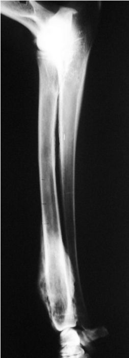
Pain was present on flexion and extension of the radio-carpal joint. Survey radiographs of the distal forelimb showed lysis of the distal radius with a sun-burst periosteal reaction and cortical
thinning (Fig. 1). A radiological diagnosis of
osteosarcoma was made. Survey thoracic radiographs (left and right laterals, dorsoventral, and ventrodorsal views)
showed no detectable pulmonary metastasis. Ultrasound examination of the left distal radius showed defects in the cortex of the radius. An ultrasound guided fineneedle aspirate was performed,
which on cytological examination confirmed the radiological diagnosis of osteosarcoma. As the dog had bilateral osteoarthrosis of the elbows secondary to elbow dysplasia and bilateral
coxarthrosis secondary to hip dysplasia, an ipsilateral vascularised ulnar transposition was done. The distal 9 cm of the left radius was excised en bloc including the involved soft tissues
while
maintaining the caudal interosseous blood vessels. A 14-hole, narrow (4.5 mm) DCP was applied to the cranial aspect of the left antebrachium and the dorsal aspect of the third metacarpal bone
(Fig. 2). Perioperative analgesia was
achieved with morphine (0.5 mg/kg every 4 hours, Morphine sulphate-Fresenius, Intramed, Port Elizabeth, South Africa) and carprofen (2.2 mg/kg bid, Rimadyl Injectable, Pfizer Laboratories,
Sandton, South Africa). The antebrachium was shortened 23 mm from a preoperative length of 222 mm, equating to a 10.4 % shortening of the limb. After the surgery the limb was placed in a modified
Robert Jones bandage. The bandage was changed
after 48 hours and then every 7–10 days. The limb was kept in a modified Robert Jones for the first 8 weeks postoperatively.
Histopathology results confirmed an osteoblastic osteosarcoma and showed incomplete excision at the caudal margin. Carboplatin (Abic Carboplatin, Teva Pharmaceuticals, Industria, South Africa) chemotherapy was started 2 days prior to surgery and repeated at 3-week intervals for 5 treatments. For each treatment thedose used was 300 mg/m2 given intravenously.
Follow-up radiographs taken on day 12 showed no loosening of the implants and on day 28 showed good callus formation, with hypertrophy of the viable ulna graft, and no implant loosening.
On day 71 the dog was presented with a weight bearing lameness of the left foreleg. Radiographs revealed clinical union of the ulna graft to the radio-carpal bone distally but not proximally. The
ulna graft had also undergone significant hypertrophy. No implant failure was detected and no radiological evidence of reoccurrence of the neoplasia was detected. Radiographs taken on day 82,
showed no pulmonary metastasis, complete clinical
union, soft tissue swelling at the caudal aspect of the antebrachium and loosening of the most proximal screw in the radius. The loose screw was subsequently removed but the client declined a
biopsy of the soft tissue swelling.
Fig. 1: Survey radiograph of the distal radius of Case 2 showing lysis of the distal radius with a sun-burst periosteal reaction and cortical thinning.
On day 126 the dog was presented with severe swelling of the distal left antebrachium, which on biopsy showed an
anaplastic malignant sarcoma (Fig. 3). On day 200 the dog was presented with a complete non-weight bearing lameness of the left foreleg and severe swelling. Thoracic radiographs were clear of
detectable metastases. A forelimb amputation including the scapula was performed. Histopathology of the prescapular lymph node revealed no neoplastic transformation. Post surgery the dog made an
uneventful recovery and coped well with the amputation. The dog was euthanased on day 464 because of severe generalised osteoarthrosis.
Fig. 2: Latero-medial and cranio-caudal radiographs of Case 1 taken immediately after limb-sparing surgery.

Fig. 3: Radiograph of Case 1 showing soft tissue swelling (thin arrows) and increased radiopacity caudal to the ulna graft. This was biopsied and proved to be local tumour recurrence. Hypertrophy of the ulna graft is clearly visible (thick arrows).

Case 2
A 10-year-old, 53 kilogram neutered male Pointer cross was presented with a histologically confirmed osteosarcoma of the left distal radius. On clinical examination pain and swelling were evident
around the left distal antebrachium.
Survey thoracic radiographs (left and right laterals, dorsoventral, and ventrodorsal views) showed no detectable pulmonary metastasis.
An ipsilateral vascularised ulnar transposition graft was performed. A narrow 4.5 mm DCP was applied to the cranial
aspect of the remaining radius and the dorsal aspect of the third metacarpal bone. Perioperative analgesia was achieved with morphine (0.5 mg/kg every 4 hours, Morphine sulphate-Fresenius,
Intramed, Port Elizabeth, South Africa)) and carprofen (2.2 mg/kg bid, Rimadyl Injectable, Pfizer Laboratories, Sandton, South Africa). The antebrachium was shortened 21 mm from a preoperative
length of 212 mm, equating to a 10 % shortening of the limb. The limb was placed in a modified Robert Jones bandage, which was changed after 48 hours and then
every 7–10 days. The limb was kept in a modified Robert Jones for the first 8 weeks postoperatively.
Carboplatin (Abic Carboplatin, Teva Pharmaceuticals, Industria, South Africa) chemotherapy was started on the day of surgery and repeated at 3-weekly intervals for 3 treatments. The dose used was 300 mg/m2 given intravenously.
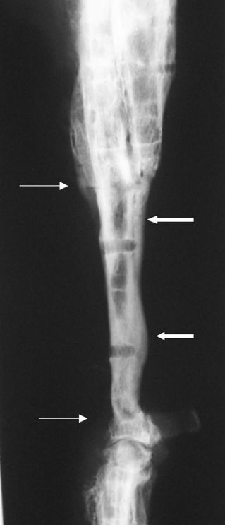
On day 16, follow-up radiographs showed that the ulna graft appeared viable but there was loosening of the screws placed in the radius as well as a longitudinal fissure in the distal end of the
radius (Fig. 4). The loose screws were
replaced with longer screws that engaged the ulna as well. The limb was maintained in a modified Robert Jones bandage.
On day 45 radiographs showed loosening of the 3 proximal screws with evidence of osteomyelitis. The loosened screws were replaced, once again engaging the ulna, and nuts were placed on the screws caudal to the ulna (Fig. 5). A swab taken from the surgical site yielded a pure growth of Pseudomonas aeruginosa. The dog was treated with ciprofloxacin (Ciploxx, Cipla Life Sciences, Belville, South Africa) at 11 mg/kg bid for 21 days.
Fig. 4: Radiograph of the proximal radius of Case 2 showing loosening of all 5 screws, with clear lysis present around all screws (black arrows).

Follow-up radiographs on day 57 showed no progression of the osteomyelitis and no loosening of the implants. By day 66 the limb function had improved significantly and thoracic radiographs detected no evidence of pulmonary metastasis. Radiographs of the limb revealed that the osteomyelitis was resolving and no implant loosening was evident. At that point new bone formation was evident between the ulnar graft and radius proximally and radio-carpal bone distally and the graft was showing hypertrophy.
Fig. 5:Case 2 following revision surgery and placement of nuts on the three proximal screws. Hypertrophy of the ulna is clearly evident.

Fig. 6: Loosening of all screws in Case 2, showing clear areas of lysis are visible around all the screws (black arrows).

Fig. 7: Case 2 following implant removal. There is complete union between the ulna and radius proximally and the ulna and radio-carpal bone distally (thin white arrows). Hypertrophy of the ulna is also clearly evident (thick white arrows).
On day 86 radiographs showed complete resolution of the osteomyelitis with no implant loosening. By day 130 function of the operated limb was excellent and thoracic radiographs were clear of metastatic disease. Clinical union of the ulna graft to the radius and radio-carpal bone was observed on radiographs taken on day 157.
On day 341 the dog was presented with acute left forelimb lameness. Radiographs showed loosening of all the screws and the distal end of the plate had lifted off the dorsal surface of the third
metacarpal bone (Fig. 6). As the ulna graft was completely fused to the radius and radiocarpal bone, the plate and all screws were removed (Fig. 7). Post-surgery limb function remained
excellent.
On day 381 the dog presented with a history of sudden episodes of pain and crying. Clinical examination revealed no
limb pain or discomfort and limb function was good. Neck pain was detected, especially on dorso-flexion and left lateral flexion. Survey radiographs of the cervical vertebrae revealed a narrowed
disc space between C5 and C6. A cisternal myelogram showed a left sided extradural compression of the spinal cord in the mid body of C6. Mild lysis was also detected in the left pedicle and left
cranial articular facet of C6. A MRI scan confirmed the myelogram
findings and was suggestive of neoplasia. The diagnostic imaging findings were confirmed on dorsal laminectomy, performed from C5 to C7. Impression smear cytology from the tissue exerting
pressure on the spinal cord showed anaplastic cells suggestive of a sarcoma. Owing to the hopeless prognosis the dog was euthanased. Histopathology confirmed a poorly differentiated sarcoma.
DISCUSSION
Osteosarcoma is the most common primary bone tumour in dogs, with the distal radius the most common site affected1,2,4,5,7,10,11. Treatment options include limb amputation or limb salvage
procedures with or without chemotherapy5. Treatment without chemotherapy is, however, seldom if ever curative, as more than 90 % of cases have micrometastases at the time of diagnosis5. Limb
amputation may not be an option in certain cases, because of musculoskeletal disease, neurological disease or client refusal. In these cases limb-sparing procedures combined
with chemotherapy would be the treatment option of choice.
Fresh frozen cortical allograft from a bone bank is the most widely used technique for limb-sparing surgery of the
distal radius. A major disadvantage of this technique is the availability, expense, and maintenance of a bone bank3,6,7,9,1
The ulnar rollover technique described in these 2 cases alleviated the need for access to a bone bank. The rollover technique has the advantage over free vascularised autograft and micro-vascular anastamosis in that it does not require specialised equipment and expertise while still having the advantage of supplying a vascularised graft10. It has an additional advantage of reduced morbidity (no distant donor site) and reduced surgical time.
The complications seen in Case 1 were local tumour recurrence and loosening of 1 screw. The loosening of the proximal screw after 82 days is not an uncommon complication13 and was addressed by
removing the screw with no further complications. Local recurrence of the tumour in this case was most likely due to incomplete tumour resection on the caudal aspect of the tumour. Attempts to
maintain vascularity of the ulna graft during surgery may have contributed to incomplete tumour removal. In retrospect the size and extent of the tumour in this case probably made this case a
poor candidate for this technique; however, the local recurrence and subsequent forelimb amputation did not compromise the overall treatment of the disease. Postsurgery the dog survived for 464
days and
was euthanased for unrelated reasons.
The complications seen in Case 2 included osteomyelitis, fracture of the proximal radius, and loosening of the
screws in the proximal radius. The loosening of the proximal radius screws was addressed by replacing the screws with longer screws, which penetrated the radius and ulna as well as utilising nuts
on the ends of the screws. The osteomyelitis resolved uneventfully once the instability was eliminated and the dog treated with antibiotics. Complete loosening of all the screws and the plate
occurred by day 341 by which time clinical union had been achieved and the implants were removed. This dog survived for 387 days following the initial surgery and was euthanased
after 387 days due to the development of, most likely, a metastatic sarcoma in the body of C6.
Clinical union was successfully achieved in both these cases, by day 82 in Case 1 and day 157 in Case 2. The delay in
Case 2 was most likely due to the implant loosening, osteomyelitis, and 2 revision surgeries. Hypertrophy of the autograft was clearly visible radiologically from an early stage in Case 1. This
indicates that vascularity of the graft was maintained and aided in strengthening the graft.
Bio-mechanically the ulna rollover technique is not as strong as a cortical allograft technique. The ulna rollover
graft was shown to fail by cranial bending of the plate in a biomechanical cadaver study10. This was not seen in either of the clinical cases described in this report. As with pancarpal
arthrodesis, 80 % or greater coverage of the metacarpal bone with the plate significantly reduces the risk of metacarpal bone fracture13. The results of the biomechanical study suggest that ulna
rollover limb-sparing cases should have external coaptation bandages for the immediate postoperative period. Both cases in this report had modified Robert-Jones bandages for a minimum of 8 weeks
postoperatively.
In these 2 cases, limb shortening did not appear to result in major mobility problems. Limb function was evaluated
subjectively by both the authors and owner and was judged to be good to excellent. The gait abnormalities seen were due to the pancarpal arthrodesis and thus mechanical in nature.
Both dogs were treated with carboplatin chemotherapy as this is a well-tolerated chemotherapeutic drug that can be given safely every 21 days at a dose of 300 mg/m2. In a study using limb amputation combined with carboplatin the median survival time was 321 days2, which is comparable with the dogs reported in this article.
Although both cases developed complications associated with the surgery, the ulna rollover technique is a technique
that can be considered for limb-sparing surgery in the dog. The risk of major infection associated with this technique is reduced to a certain extent and the ability to resolve any infection is
also increased by the vascularity of the graft. The most significant potential problem in our opinion is that of complete tumour excision. This technique is therefore best used for those cases
with small tumour size that have not broken through the cortex, especially caudally.
REFERENCES
1. Berg J 1996 Canine osteosarcoma – amputa-
tion and chemotherapy. Veterinary Clinics of
North America: Small Animal Practice 26:
111–121
2. Bergman P J, MacEwen E G, Kurzman I D,
Henry C J, Hammer A S, Knapp D W, Hale
A, Kruth S A, Klein M K, Klausner J, Norris
A M, McCaw D, Straw R C, Withrow S J 1996
Amputation and carboplatin for treatment
of dogs with osteosarcoma: 48 cases (1991 to
1993). Journal of Veterinary Internal Medicine
10: 76–81
3. Buracco P 2002 Pasteurised tumoural
autograft as a novel procedure for limb
sparing in the dog: a clinical report. Veteri-
nary Surgery 31: 525–532
4. Dernell W S 2003 Limb-sparing surgery for
dogs with bone neoplasia. In Slatter D (ed.)
Textbook of small animal surgery Vol. 2 (3rd
edn). W B Saunders, Philadelphia: 2272–
2284
5. Garzotto C 2003 Canine appendicular
osteosarcoma. In Slatter D (ed.) Textbook of
small animal surgery Vol. 2. (3rd edn). WB
Saunders, Philadelphia: 2460–2474
6. Liptak J M, Dernell W S, Ehrhart N,
Withrow S J 2004 Canine appendicular
osteosarcoma: diagnosis and palliative
treatment. Compendium on Continuing
Education for the Practicing Veterinarian 26:
172–182
7. Liptak J M, Dernell W S, Ehrhart N,
Withrow S J 2004 Canine appendicular
osteosarcoma: Curative intent treatment.
Compendium on Continuing Education for the
Practicing Veterinarian 26: 182–196
8. MacEwen E G 1996. Canine osteosarcoma:
Amputation and chemo-immunotherapy.
Veterinary Clinics of North America: Small
Animal Practice 26: 123–133
9. Morello E 2003 Pasteurised tumoral auto-
graft and adjuvant chemotherapy for the
treatment of canine distal radial osteosar-
coma: 13 cases. Veterinary Surgery 32: 539–
544
10. Pooya H A, Séguin B, Mason D R, Walsh P J,
Taylor K T, Kass P H, Stover S M 2004 Bio-
mechanical comparison of cortical radial
graft versus ulnar transposition graft limb-
sparing techniques for the distal radial site
in dogs. Veterinary Surgery 33: 301–308
11. Sequin B 2003 Use of an ipsilateral vascu-
larised ulna transposition autograft for
limb-sparing surgery of the distal radius in
dogs: an anatomic and clinical study. Veteri-
nary Surgery 32: 69–79
12. Straw R C 1996 Limb-sparing surgery versus
amputation for dogs with bone tumors. Vet-
erinary Clinics of North America: Small Animal
Practice 26: 135–143
13. Whitelock R G, Dyce J, HoultonJEF 1999
Metacarpal fractures associated with pancarpal
arthrodesis in dogs. Veterinary Surgery 28: 25–30
Share this article / Teilen Sie diesen Artikel